

What is a dental microscope and what does it change in dental treatment?
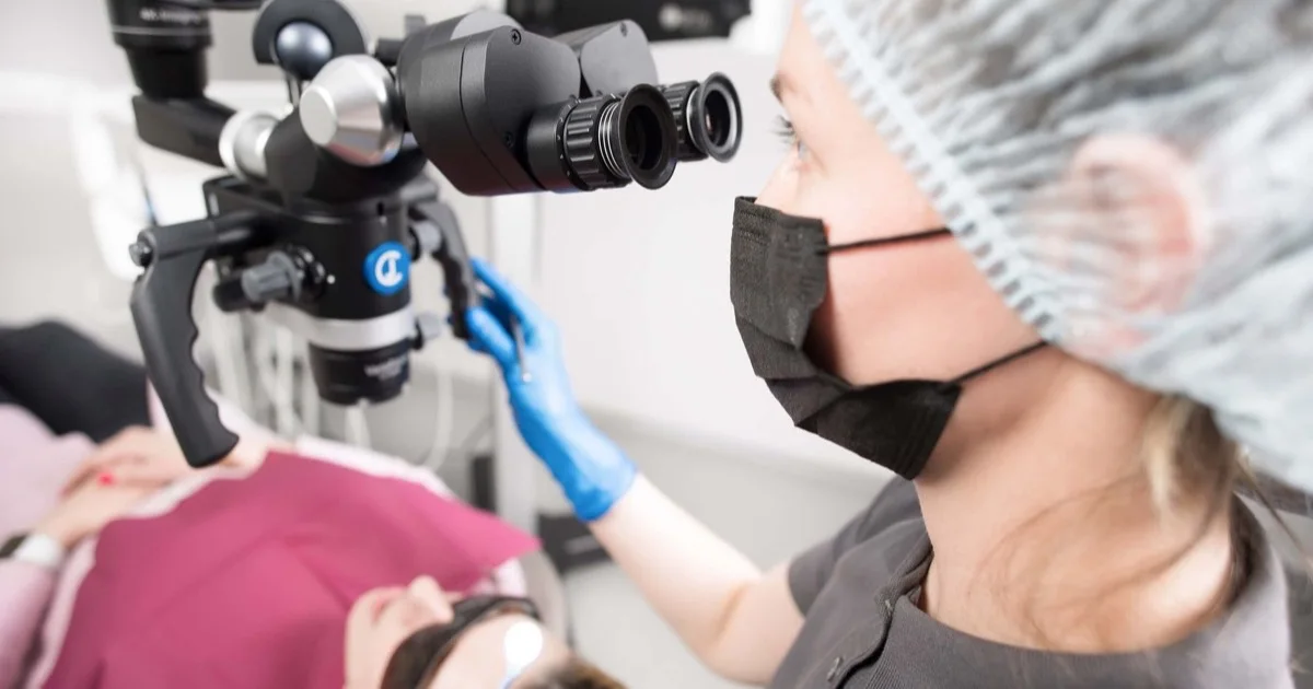
A dental microscope (surgical microscope) is a device that magnifies the working area many times over and lights it up with a powerful beam: extra root canals, micro-cracks, remnants of old filling material and the margins of a restoration that the naked eye cannot pick out become visible. Its greatest benefit comes in root canal treatment and dental surgery: the dentist works by seeing, not by feeling. In the scientific literature, the modern technique of apical surgery performed under magnification is associated with a significantly higher success rate than the traditional method.
What is a dental microscope?
It is an optical device mounted above the dental chair: an objective lens, magnification steps (usually from around 4× up to 20× and beyond) and a powerful LED light focused on the working area. The dentist looks through the eyepieces of the microscope throughout the treatment and can change the magnification at any moment. At Smile by Dr. Bakhtiyar, we use a German-made CJ-Optik surgical microscope.
Two things set a microscope apart from loupes: the magnification is many times greater, and the beam of light falls directly inside the working area — even the floor of a deep canal appears well lit.
How is it used?
- Diagnostics. An area that looks suspicious on an X-ray — a crack, hidden decay, a gap at the margin of an old filling — is checked directly under magnification.
- In root canal treatment. Canal orifices are located under magnification: an extra canal often found in upper molars (for example, MB2), curved or narrowed canals, an instrument fractured inside a canal — all of these come into the field of view. Cleaning and filling are also carried out under visual control.
- In surgery. During apical surgery, sinus lift and implant placement it allows work through small incisions, with minimal intervention in the tissues.
- In restorations. Precise finishing of filling and crown margins is one of the main ways of reducing marginal gaps.
Which treatments do we use it for?
- Root canal treatment — for both initial treatment and retreatment (revision); we have written separately about why it is painless: is root canal treatment painful?
- Dental surgery — apical surgery and minimally invasive procedures.
- Treatment of tooth decay and restorations — selective removal of tissue in deep decay, marginal precision.
- Assessment of inflammatory conditions — where the source of the symptoms is unclear; see: dental abscess and signs of infection.
What does it mean for the patient?
- Less healthy tissue touched — the dentist works only on the affected area, because it can be seen precisely.
- Hidden problems are found — an extra canal that goes undetected is a common cause of future infection; magnification helps to reduce this risk.
- A more predictable result — a canal filled under visual control and precisely finished margins serve the longevity of the work.
- Documentation — with a camera attached to the microscope, photo and video records of the working area can be taken; you can also see what is happening inside.
What does the scientific evidence say?
The strongest evidence comes from surgical endodontics. In the meta-analysis by Setzer and colleagues (Journal of Endodontics), traditional apical surgery was compared with microsurgery: the success rate was significantly higher with microsurgery, carried out using a microscope, microinstruments and modern filling materials. A second meta-analysis by the same group compared the technique with and without magnification and showed that high magnification gives an advantage, particularly in posterior teeth.
An honest note: for conventional (non-surgical) root canal treatment, the evidence on how much the microscope directly changes the outcome is more limited — here the microscope is first and foremost a tool for visualisation, precision and finding hidden anatomy, while success still depends on many factors (the condition of the tooth, isolation, the quality of the filling, the restoration). The microscope does not replace a good protocol — it strengthens it.
How is the microscope used at Smile by Dr. Bakhtiyar?
At our clinic in Baku, the CJ-Optik microscope is a standard part of root canal treatment and minimally invasive surgery: the search for canal orifices, the removal of old material during retreatment and work at the root tip are all carried out under magnification. The microscope works together with rubber dam isolation and digital diagnostics — the full list of equipment is on the Technology page.
Frequently asked questions
Does treatment with a microscope take longer? The appointment may be somewhat longer, because the work is more detailed. In return, finding hidden problems at that very first appointment reduces the likelihood of retreatment later on.
Is a microscope needed for every root canal treatment? In cases with simple anatomy, treatment can be carried out to a high standard without a microscope as well. In curved, narrow canals, cases with extra canals and those requiring retreatment, the benefit of magnification is greater; the dentist makes the decision.
Is there any extra discomfort for the patient? No. The microscope only changes what the dentist sees; for you the process is no different from ordinary treatment.
Does a microscope replace X-rays? No. X-rays and CBCT show the inside of the tooth and the bone, while the microscope magnifies the open working area — these are tools that complement one another.
Can a fractured instrument be removed with a microscope? In many cases, yes — being able to see the instrument greatly increases the chances of removing it. Each specific situation is assessed during the examination.
Clinical note: This article is for general information only and does not replace individual advice from your dentist. To discuss your plan for root canal treatment or a surgical procedure, book an appointment.
Sources
- Outcome of endodontic surgery: a meta-analysis of the literature — Part 1: Comparison of traditional root-end surgery and endodontic microsurgery — Journal of Endodontics, 2010 (Setzer et al.)
- Outcome of endodontic surgery: a meta-analysis of the literature — Part 2: Comparison of endodontic microsurgical techniques with and without the use of higher magnification — Journal of Endodontics, 2012 (Setzer et al.)
- CJ-Optik — dental surgical microscopes — CJ-Optik GmbH & Co. KG (istehsalçı), Almaniya
This article is for general information only and does not replace an individual diagnosis, examination or treatment. Consult a doctor for a decision about your case.