

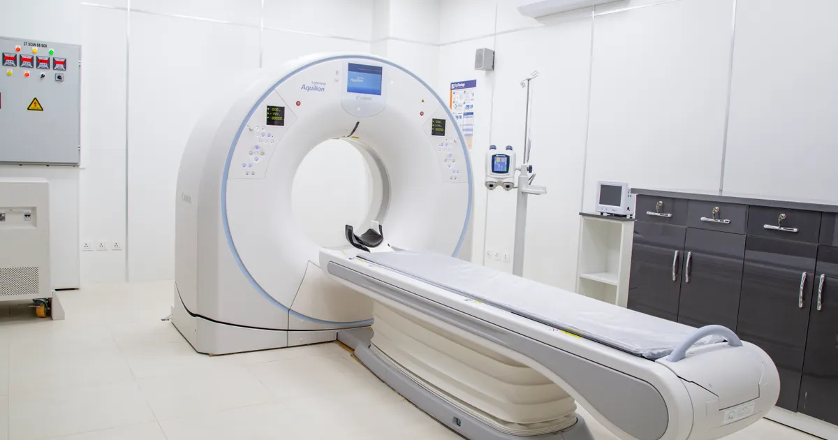
CBCT (Cone Beam Computed Tomography) is a special dental X-ray method that uses a cone-shaped beam to show the jaws, teeth and facial bones in three dimensions (3D). Unlike conventional CT, a single rotation (~15–25 seconds) produces a 3D model of the whole area, while the radiation dose is considerably lower than with medical CT.
At the clinic, CBCT is taken when indicated and with the smallest suitable field of view — the workflow for this service is set out on the 3D diagnostics page.
In modern dentistry, CBCT is considered the gold standard for implant planning, root canal treatment, orthodontics and jaw surgery. In this article we explain it from a dentist's point of view: how the machine works, when it is useful, what the dose is and what the procedure is like for the patient.
What does CBCT mean and what does the abbreviation stand for?
CBCT is short for Cone Beam Computed Tomography. In Azerbaijani it is translated as konus-şüalı kompüter tomoqrafiyası (KLKT) or simply 3D dental tomography or 3D diagnostics. Many patients know it as a 3D jaw scan or a 3D dental X-ray — these are all the same examination.
On a conventional (2D) dental X-ray, structures overlap and depth is lost. CBCT, on the other hand, combines many images to create a volumetric model that can be sectioned and viewed from any angle. This means the dentist can look at the same image from the side, from the front, from above and at any layer; measure the thickness and height of the bone; and see the exact position of the nerve canal and the nasal cavity.
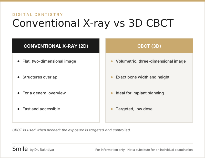 Conventional X-ray versus 3D CBCT: the main differences.
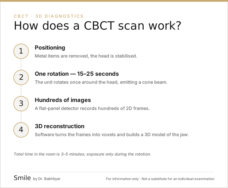
How does CBCT work? — A simple explanation
A CBCT machine has three main parts: an X-ray source, a flat panel detector and a rotating arm. The procedure goes like this:
- The patient sits inside the machine or stands, keeping the head still.
- The machine makes 1 rotation around the head (usually 15–25 seconds).
- During this time the X-ray source emits a cone-shaped beam and the detector records hundreds of two-dimensional images.
- Special reconstruction software converts these images into three-dimensional pixels called voxels and creates a precise 3D model of the jaw.
As a result, the dentist can section the jaw in any plane — axial (from above), sagittal (from the side) and coronal (from the front) — and measure with micron accuracy.
Why does the cone-shaped beam matter?
A conventional medical CT (spiral / MSCT) emits a flat, fan-shaped beam and images the jaw as hundreds of thin slices. That means more time and more radiation. CBCT covers the whole area with a single cone beam — faster, a lower dose, while keeping the accuracy needed for teeth. You can read more about these differences in our article The difference between CBCT and conventional CT.
Which areas does CBCT show?
In a single CBCT scan the dentist can see all of the following at once:
- All the teeth — including roots, crowns and unerupted teeth.
- The lower and upper jawbones — density, height, width.
- The sinus cavities (maxillary sinuses) — inflammation, thickening of the membrane.
- The lower jaw nerve (mandibular canal) — an essential guide for implants.
- The temporomandibular joint (TMJ) — the position of the condyle and structural disorders.
- The nasal cavity and surrounding bones.
The images are stored in DICOM format and can be imported into any planning software (for example, implant planning programs).
The most common uses of CBCT
Not every patient needs a CBCT — it is taken when there is an indication. It is most often recommended in the following situations:
- Dental implant planning — to measure bone height, width and density precisely.
- Difficult root canal treatment — to see extra canals and root curvature.
- Wisdom tooth removal — to assess how close the tooth is to the lower jaw nerve.
- Orthodontic treatment — the position of impacted teeth (teeth remaining under the gum) and the condition of the roots.
- TMJ problems — pain or clicking when opening and closing the mouth.
- Toothache of sinus origin — the relationship between the upper teeth and the sinus.
- Trauma and jaw surgery — assessment of fractures, cysts and tumours.
To find out whether CBCT is really necessary for a particular treatment, you can read our article When CBCT is essential.
Radiation dose: is CBCT safe?
This is the question that worries patients most. The short answer: a CBCT taken for the right indication is safe, because the dose is very low.
Approximate comparison (in microsieverts, µSv):
- One flight (7 hours, atmospheric radiation): ~40 µSv
- A conventional periapical (single tooth) X-ray: ~5 µSv
- Panoramic X-ray: ~15–25 µSv
- Dental CBCT (small FOV): ~40–100 µSv
- CBCT (large FOV, full jaw): ~100–200 µSv
- Standard medical head CT (MSCT): ~1000–2000 µSv
In other words, a typical dental CBCT delivers a dose 5–20 times lower than a medical CT and is equivalent to a few weeks of everyday natural background radiation (in a 2015 meta-analysis, with standard protocols the average dose in adults was 84 µSv for a small FOV and 212 µSv for a large FOV; the differences between machines are considerable). Even so, the principle remains: the dose must always be justified (ALARA — as low as reasonably achievable). International guidelines (the ADA's 2024 recommendations, the European SEDENTEXCT document) recommend taking a CBCT only when 2D X-rays are not sufficient. If you are pregnant or have any other special condition, be sure to tell your dentist before the scan. More on this: Is CBCT radiation high?.
A special rule for children
Cells in children are more active, so they are more sensitive to radiation. For this reason a CBCT in children is only taken when there is a clear indication, and with the smallest possible FOV (field of view).
CBCT is the foundation of digital planning
The volumetric model obtained from CBCT combines with the other stages of digital dentistry: it is superimposed on the intraoral scan, the implant is positioned virtually in the software, a surgical guide (template) is made if needed, and the treatment is planned in advance. For the patient this means three practical differences:
- fewer surprises — the plan is built before the procedure, so uncertainties are reduced;
- clearer conversations — the dentist can show the situation on screen in cross-sections;
- more precise stages — the sequence and duration of treatment are known in advance.
For how this workflow works for implants: Why CBCT matters for dental implants.
What is the CBCT procedure like for the patient?
In practice the procedure is very simple:
- The patient removes earrings, hairpins, dentures and glasses.
- The neck and chest area are protected with a lead apron.
- The patient steps up to the machine, the chin is placed on the chin rest and the head is stabilised with a fixator.
- The dentist or radiographer advises: please do not move and do not swallow.
- The machine makes a 15–25 second rotation; radiation is emitted only during this short period.
- The total time spent in the room is 3–5 minutes.
The procedure is painless, no contrast agent is used, and no special preparation or fasting is required. It is also comfortable for patients with claustrophobia, because CBCT is an open machine — the patient does not go inside a tunnel.
How is the CBCT result delivered?
Within 5–15 minutes of the scan the dentist receives the following:
- A DICOM file — the raw file holding all the 3D data, which can be shared on a USB stick or via the cloud.
- A PDF report — with the main cross-sections, a panoramic reconstruction and explanations.
- Images for planning — for implant, orthodontic or surgical plans.
Thanks to the DICOM file, dentists at different clinics can work with the same data. For more on how the result is read, see our article How a CBCT result is read.
How long does a CBCT scan take?
The radiation part alone takes 15–25 seconds (20–35 seconds for a large field of view, 10–15 seconds in the small endodontic mode). Including walking in and out of the room, the total time is usually 3–5 minutes. With children and anxious patients, positioning may take a little longer.
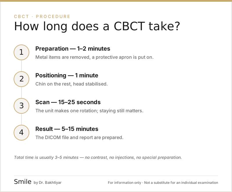
Step by step:
- Preparation (30–60 seconds) — earrings, necklaces, glasses and removable dentures are taken off; a protective apron is put on.
- Positioning (30–60 seconds) — the chin is placed on the rest, the forehead is fixed, and the plane is selected using light guides.
- Exposure (15–25 seconds) — the machine rotates once around the jaw; you are asked not to move and not to swallow; there is no pain or discomfort.
- Reconstruction (30 seconds – 3 minutes) — the software converts the image into 3D voxels; the DICOM file and the first images are ready within 5–15 minutes.
At our clinic the raw DICOM file and the first images are provided during the same visit, and the dentist's written explanation is usually given the same day; the treatment plan is discussed at the next appointment. For more on how the result is read, see our article How a CBCT result is read.
FAQ — Frequently asked questions
1. Are CBCT and a panoramic X-ray the same thing?
No. A panoramic X-ray shows the teeth on a single flat sheet (2D). CBCT is 3D — it gives depth, density and precise measurements. A panoramic X-ray is for an initial overview, CBCT is for precise planning.
2. Can a CBCT be taken during pregnancy?
Usually not. During pregnancy any planned X-ray is postponed. It is taken only on the dentist's decision, in cases of urgent medical indication and with protective measures in place.
3. Can patients with a pacemaker or an implant have a CBCT?
Yes. CBCT does not use a magnetic field (it should not be confused with MRI), so it is not a problem for pacemakers or metal implants. Metal may only create a small artefact on the image.
4. How often can a CBCT be taken?
When there is a clinical indication. It is not for routine monitoring — it is repeated every 1–3 years only when there is a new treatment plan or a noticeable change.
5. In what format is the CBCT result provided?
DICOM (the raw 3D file) and a PDF report. The patient can receive it both on a USB stick and via a cloud link, and can share it with another dentist.
6. If I have older X-rays, do I need a new scan?
The dentist assesses the existing images. For implant and surgical planning a three-dimensional image is often required, because a conventional X-ray does not show the volume of the bone; if there is a recent enough CBCT covering the area needed, a repeat scan is not necessary.
Conclusion and booking an appointment
CBCT is a technology that makes decision-making in modern dentistry considerably easier in implant treatment, root canal treatment, orthodontics and jaw surgery. Used for the right indication, it is low-dose and, for the patient, a painless 3–5 minute procedure.
If you are planning a dental implant, wisdom tooth removal, root canal treatment or orthodontics, 3D imaging can fundamentally improve the quality of your treatment. Our clinic in Baku has a modern CBCT machine and the expertise needed to read the results.
👉 Book an appointment for dental implants and our other services or get in touch via WhatsApp.
Author: Dr. Bakhtiyar Aliyev — dentist and head dentist of the clinic. Specialised in aesthetic and implant dentistry and jaw surgery. This article has been reviewed by a dentist.
Sources
- Effective dose of dental CBCT — a meta analysis of published data and additional data for nine CBCT units — Dentomaxillofacial Radiology, 2015
- Optimizing radiation safety in dentistry: Clinical recommendations and regulatory considerations — Journal of the American Dental Association, 2024
This article is for general information only and does not replace an individual diagnosis, examination or treatment. Consult a doctor for a decision about your case.