

3D Diagnostics
3D CBCT diagnostics at Smile by Dr. Bakhtiyar in Baku shows your teeth, jawbone and nerve canals in three dimensions — the base of precise treatment.
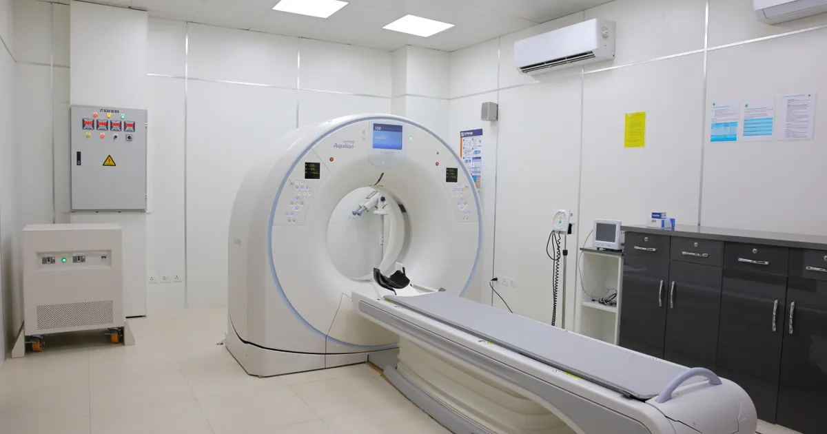
3D diagnostics means cone beam computed tomography (CBCT) — a scan that shows your teeth, jawbone, sinuses (air spaces near the nose) and nerve canals in three dimensions. At Smile by Dr. Bakhtiyar in Baku this scan is the base of our digital planning. A normal X-ray shows everything on one flat image, while CBCT lets the dentist see the jaw from every side, in real size.
A 3D scan is most often needed before an implant, to measure the height and width of the bone. It also helps with an impacted wisdom tooth, sinus lift planning, difficult root canals, pain that does not go away, the search for cysts or hidden inflammation, orthodontic planning and jaw joint problems.
The process is simple. First you remove earrings, glasses, removable dentures and other metal items. Then you stand or sit at the scanner and rest your chin on the support. The machine makes one quiet turn around your head and the scan takes only a few seconds. There is no pain at all. The image appears on the computer at once and your dentist explains it on the screen.
There are no limits after the scan — you can go straight back to work and your normal day. The result is stored as a digital file and used to build your treatment plan. Radiation is kept low and the scan is only prescribed when it is really needed. Please tell your dentist before the scan if you are pregnant or have any concerns.
A 3D tomography costs 100 AZN, a panoramic X-ray 30 AZN, and the first visit with consultation and a personal treatment plan 60 AZN. The scan itself takes a few minutes and the results are usually reviewed during the same visit. Your dentist decides after the examination whether a 3D scan is needed — it is not required in every case.
What is 3D diagnostics (CBCT)?
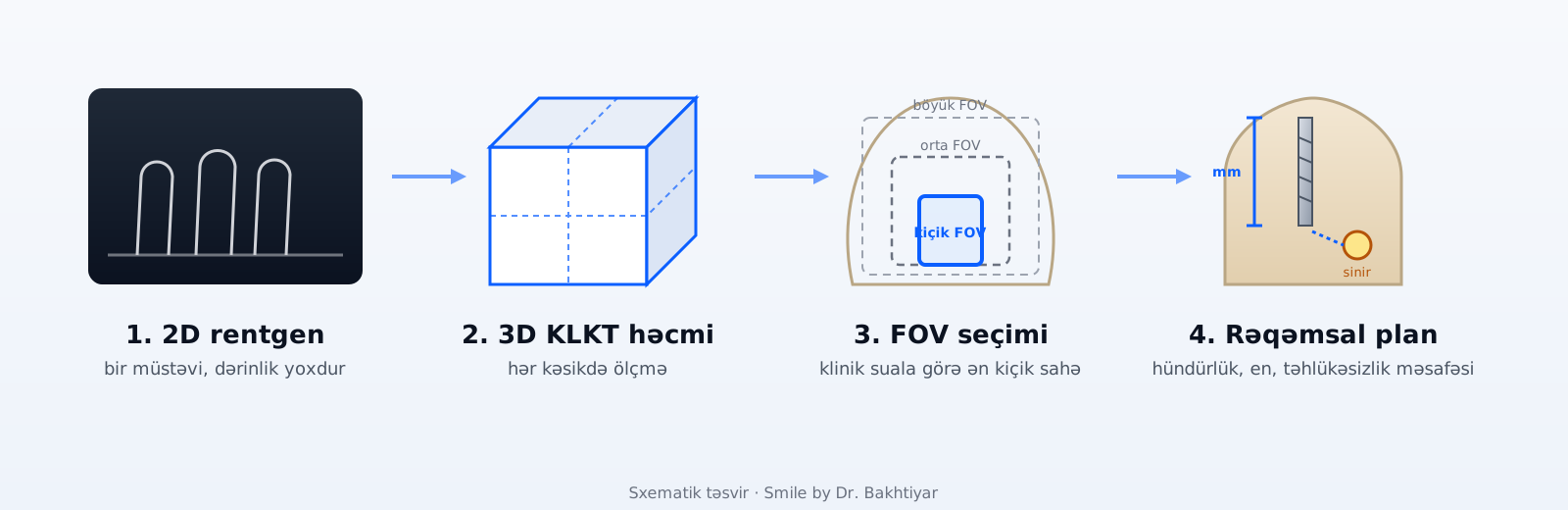
Cone-beam computed tomography (CBCT) is an X-ray method that captures a three-dimensional volume of the jaws, teeth, sinuses and jaw joint in a single rotation. A conventional X-ray compresses structures into one plane; CBCT lets the dentist measure height, width and depth in any slice. In the clinic CBCT is not a routine examination — it is an indication-based service that answers a specific clinical question in implant, endodontic, surgical and orthodontic planning.
When is it used?
The general rule: not “3D for every patient”, but 3D when 2D cannot answer the question.
- Implant planning — bone height and width, distance to the nerve canal and sinus (the 2012 AAOMR position recommends cross-sectional imaging)
- Endodontics — complex canal anatomy, a persistent periapical lesion, suspected root fracture, retreatment planning
- Wisdom teeth — the root's relationship to the nerve canal when the panoramic X-ray shows risk signs
- Cysts, tumours and bone pathology — size, borders, neighbouring structures
- Jaw joint — bony changes of the condyle, trauma
- Orthodontics — impacted teeth, suspected root resorption, skeletal asymmetry (AAOMR 2013: not routine, on indication)
- Sinus — odontogenic sinusitis and sinus-lift planning
Its role in implant planning
The implant's length, diameter and angle are chosen from the real bone dimensions; the safety distance to the nerve canal in the lower jaw and to the sinus in the upper jaw is measured on the slices. According to a systematic review (2014) and the AAOMR position, cross-sectional imaging of the implant site is the standard recommendation; CBCT data is merged with the intraoral scan and the design of the future tooth to produce the digital plan and, where needed, a surgical guide. Bone deficiency (the need for grafting or a sinus lift) is identified here as well.
Its role in endodontics
According to the AAE/AAOMR joint position statement (2015), CBCT is not routine in root canal treatment; a limited (small-FOV) scan is indicated for complex anatomy, an unlocated canal, persistent symptoms, suspected root fracture or resorption and surgical endodontic planning. A 2026 meta-analysis confirms that CBCT detects periapical lesions significantly more often than 2D radiographs — but not every detected change needs treatment; the decision is made together with the clinical picture.
Its role for wisdom teeth
The first step in assessing a wisdom tooth is a panoramic X-ray. When risk signs are present — the root overlapping the nerve canal, darkening or narrowing of the canal — CBCT shows the root's position relative to the canal (buccal/lingual, contact) and can change the surgical plan. Without risk signs CBCT is usually not needed.
Its role in jaw-joint disorders
Most jaw-joint complaints are diagnosed clinically. CBCT is used on indication to show the bony components — condyle shape, cortical bone, degenerative changes, trauma; according to a systematic review (2024) it is highly accurate for detecting bony changes, while MRI is preferred for the disc and soft tissues.
Its role in orthodontics
According to the 2013 AAOMR recommendations, CBCT is not a routine orthodontic record; it is indicated for impacted and abnormally positioned teeth, suspected root resorption, clefts and skeletal asymmetry and mini-screw planning — with the smallest appropriate FOV. Panoramic and cephalometric X-rays remain the standard orthodontic records.
What is FOV?
FOV (field of view) is the scanned volume. A small FOV (a few teeth, for example for root canal treatment) gives higher resolution and a lower dose; a medium FOV covers one jaw, a large FOV both jaws, the sinuses and the joints. Dose and interpretation burden increase with FOV, so the dentist chooses the smallest field that covers the clinical question — the practical application of the ALADA principle (“as low as diagnostically acceptable”).
How is the scan performed?
- 1
Indication and registration
The treating dentist defines the clinical question and the region needed; pregnancy, previous images and metal objects are asked about.
- 2
Preparation
Earrings, necklaces, glasses and removable dentures are taken off; a protective apron is put on.
- 3
Positioning
The chin is placed on the rest, the head is stabilised, and the correct plane is set with light guides.
- 4
FOV and protocol selection
According to the indication, the smallest appropriate field of view, voxel size and dose mode (child/adult) are selected.
- 5
Scan
The unit makes one rotation around the head; exposure lasts 10–35 seconds; you need to stay still, there is no pain.
- 6
Reconstruction
The software converts the volume into 3D voxels; the DICOM file and slices are ready in a few minutes.
- 7
Clinician interpretation and plan
The image is interpreted together with the clinical data; measurements and findings go into the treatment plan and are explained to the patient on screen.
How long does the scan take?
The exposure itself is 10–35 seconds and the total time in the room about 3–5 minutes; the DICOM file and initial images are given at the same visit, and the dentist's written interpretation is usually ready the same day. A stage-by-stage timeline is in the blog article.
Radiation dose
The dose is not a universal number — it depends on the field of view, the device and the protocol. According to a 2015 meta-analysis, the effective dose of dental CBCT averages ~34 μSv for a small FOV, ~88 μSv for a medium and ~131 μSv for a large one; for comparison, a panoramic X-ray is a few tens of μSv and a medical head CT usually 1–2 mSv. Children are more radiosensitive, so a paediatric mode and a small FOV matter. The clinic's approach is indication-based: the scan is ordered only when a question could change the outcome, the smallest appropriate FOV is selected, and an existing recent image is not repeated. The phrases “completely safe” or “needed for everyone” are not used — benefit and risk are assessed for each patient individually.
CBCT versus medical CT
CBCT collects the volume in one rotation with a cone beam and a flat-panel detector and gives high resolution for bone and teeth; medical CT (MSCT) is superior in soft-tissue contrast and density measurement, and its dose is usually higher. For questions about teeth and jawbone the clinic chooses CBCT; for tumours, spread of infection into soft tissue or questions about the joint disc the patient is referred for medical CT/MRI.
How are the results used?
A CBCT image is data, not a “report”: the treating dentist interprets it together with the complaint, the examination and other X-rays. In implant planning the measurements become the digital design, in endodontics a canal map, in surgery an access plan. Systematic reviews show that incidental findings on CBCT (sinus changes, bone islands, impacted teeth) are common — not all are disease, but they are recorded and a radiologist's report is obtained when needed. The DICOM file is given to the patient.
Risks and limitations
- Radiation — low, but not zero; a scan without indication is not justified
- Soft tissue — limited contrast; MRI/CT for the disc, muscles and mucosa
- Metal artefacts — image quality can drop around crowns, implants and fillings
- Motion artefacts — in children and anxious patients
- Incidental findings — require additional interpretation and sometimes further investigation
- Over-diagnosis — not every detected change needs treatment
- Density measurement — CBCT does not provide reliable Hounsfield units
Price
The prices below come from the same source as the site's Prices section. CBCT can be taken as part of treatment (implant, endodontic or surgical planning) or separately on referral from another dentist; in either case the patient receives the DICOM file and the images.
| Initial visit, consultation and personalised treatment plan | 60 AZN |
| Panoramic X-ray | 30 AZN |
| 3D tomography (CBCT) | 100 AZN |
Prices come from the same source as the Prices page; the final amount is set after your examination. All prices →
Doctor and clinic
At Smile by Dr. Bakhtiyar (Baku, Babek Avenue 1131) the indication for CBCT and its interpretation are decided by the treating dentist; implant and surgical planning is led by Dr. Bakhtiyar Aliyev, orthodontic and jaw-joint questions by Dr. Maharram Mammadov. The image forms the basis of the clinic's digital planning workflow — intraoral scan, design, surgical guide.
Sources
- Position statement of the American Academy of Oral and Maxillofacial Radiology on selection criteria for the use of radiology in dental implantology with emphasis on cone beam computed tomography — Oral Surgery, Oral Medicine, Oral Pathology and Oral Radiology, 2012 (AAOMR)
- AAE and AAOMR Joint Position Statement: Use of Cone Beam Computed Tomography in Endodontics — 2015 Update — Oral Surgery, Oral Medicine, Oral Pathology and Oral Radiology, 2015 (AAE/AAOMR)
- Clinical recommendations regarding use of cone beam computed tomography in orthodontics. Position statement by the American Academy of Oral and Maxillofacial Radiology — Oral Surgery, Oral Medicine, Oral Pathology and Oral Radiology, 2013 (AAOMR)
- Cone beam computed tomography in implant dentistry: a systematic review focusing on guidelines, indications, and radiation dose risks — International Journal of Oral & Maxillofacial Implants, 2014
- Effective dose of dental CBCT — a meta analysis of published data and additional data for nine CBCT units — Dentomaxillofacial Radiology, 2015
- Diagnostic Accuracy of CBCT Versus 2D Radiography for Various Dental Pathologies: A Systematic Review and Meta-Analysis — Australian Endodontic Journal, 2026
- The diagnostic accuracy of cone beam computed tomography in detecting temporomandibular joint bony disorders: a systematic review — Polish Journal of Radiology, 2024
- Incidental findings from cone-beam computed tomography in children and adolescents: a systematic review — European Archives of Paediatric Dentistry, 2025
If this sounds familiar
If you have one of these complaints, this service may be right for you:
- “I want an implant but I don't know if I have enough bone”
- “My wisdom tooth never came out, they say it is in the bone”
- “The tooth was removed but the pain is still there”
- “Something shows on my X-ray and I want to know exactly”
- “An upper tooth was taken out and they mention a sinus problem”
- “My root canal was treated but the swelling comes back”
- “My jaw clicks and my mouth does not open fully”
- “There is a swelling on my gum that never goes away”
Frequently asked questions
When is a 3D scan (CBCT) needed?
When there is a specific clinical question: bone height/width and the relationship to the nerve and sinus for an implant, complex canal anatomy or a persistent periapical problem, a wisdom tooth close to the nerve, a suspected cyst or tumour, bony changes of the jaw joint, impacted teeth. It is not ordered for routine check-ups.
Isn't a conventional X-ray enough?
In many cases it is — panoramic and intraoral X-rays remain the first choice. CBCT is added only where 2D cannot answer the question: depth, bone width, the exact position of the nerve, the three-dimensional relationship of a root.
What is the radiation dose of CBCT?
It depends on the field of view (FOV), the device and the protocol: a meta-analysis reports on average ~34 μSv for a small FOV, ~88 for medium and ~131 for large; a medical head CT is usually 1–2 mSv. The clinic selects the smallest appropriate FOV and an indication-based protocol (ALADA principle); “zero dose” or “completely safe” are not correct statements.
How long does the scan take and does it hurt?
Exposure lasts 10–35 seconds, total time in the room 3–5 minutes; there is no pain or discomfort, you only need to stay still. A step-by-step description is in the blog article.
Can CBCT be done during pregnancy?
During pregnancy imaging is performed only for urgent indications and with protective measures; elective scans are usually postponed until after delivery. Tell the dentist about pregnancy or a suspected pregnancy before the scan.
Is CBCT used in children?
Only for a specific indication (impacted tooth, trauma, anomaly) and with the smallest FOV; because children are more radiosensitive, 2D methods are considered first. Incidental findings are also common in children and need interpretation.
What is FOV and why does it matter?
Field of view — the scanned volume. A small FOV (a few teeth) gives higher resolution and a lower dose; a large FOV (both jaws, sinuses, joints) is for broad planning. The dentist chooses the smallest FOV that covers the question.
Who interprets the result, and can I take it with me?
The treating dentist interprets the image together with the clinical findings; for an unusual finding a radiologist's report is obtained. The DICOM file and images are given to the patient — they can be used in another clinic or for a second opinion.
Does CBCT show soft tissue?
Only to a limited extent — CBCT is optimised for bone and teeth; for details of the gums, muscles, joint disc or sinus lining MRI or medical CT may be needed. This is one of the method's main limitations.

Doctor performing this treatment
Implantologist · Digital Dentistry · Founder of the clinic