

What is FOV and how is it chosen?
FOV (Field of View — the imaging area) is the size of the cylindrical volume that a CBCT machine captures as an image in a single scan. It is usually given as diameter × height (e.g. 5×5 cm, 8×8 cm, 15×15 cm). A small FOV covers only one specific region (one or two teeth, a single sinus, a single jaw joint) and keeps the dose to a minimum. A large FOV, on the other hand, shows the entire jaw and facial structures, but requires more radiation. Choosing the right FOV means selecting the smallest area required by the clinical indication; this is in line with the ALARA (As Low As Reasonably Achievable) principle.
Choosing the FOV is part of the protocol stage of our 3D imaging service.
Why does FOV matter?
In CBCT, two key technical parameters determine both the quality of the information and the dose:
- FOV (field of view): the larger it is, the more anatomy is captured, but the dose increases and the voxel size is on average larger (small details may be lost).
- Voxel size (image resolution): a smaller voxel means a more detailed image, but scan time and dose may increase.
Choosing the wrong FOV creates two problems:
- Too small a FOV: the area around the clinical problem is not visible and a second scan is needed → the patient receives a double dose.
- Too large a FOV: areas that are not needed are also scanned → unused radiation, excess dose.
Striking this balance is the professional responsibility of the dentist who requests the CBCT.

FOV categories
Dental CBCT machines usually offer three FOV categories:
1. Small FOV (~4×4 cm – ~6×6 cm)
For a single region only: one or two teeth, the root and periapical area, a single sinus floor, one jaw joint.
Indications:
- Endodontic (root canal) diagnosis — suspected inflammation at the apex of a tooth with previous root canal treatment;
- Root fracture and resorption;
- Complex root anatomy before a tooth extraction;
- Planning a single implant site;
- Assessment of one TMJ.
Advantage: the lowest dose (0.02–0.08 mSv), the highest detail (small voxel).
2. Medium FOV (~8×8 cm – ~10×10 cm)
For half or a full arch: the posterior region of one jaw, the sinuses, the entire alveolar ridge.
Indications:
- Planning several implants;
- Sinus lift assessment;
- Locating orthodontically displaced teeth;
- Surgical wisdom tooth extraction (relationship with the lower jaw nerve);
- Assessment of cysts and tumours.
Dose: approximately 0.05–0.15 mSv (depending on the machine).
3. Large FOV (~13×15 cm and above)
For the whole maxillofacial region (upper jaw + lower jaw + condyle + nose).
Indications:
- Orthognathic surgery planning;
- Complex orthodontic (skeletal Class II/III) treatment planning;
- Craniofacial anomalies (cleft palate, etc.);
- Multilocular, widespread cysts and tumours;
- Bilateral TMJ pathology.
Dose: approximately 0.1–0.3 mSv.
Clinical principles of FOV selection
The recommendations of international radiological societies (SEDENTEXCT, EAO, AAOMR) are as follows:
- The smallest FOV is chosen according to the clinical indication — the ALARA principle;
- The ROI (Region of Interest) must be fully covered in the axial plane — the area around the clinical problem should be visible with a 5–10 mm margin;
- A fine voxel must be balanced against clinical need — a 100–150 µm voxel for endodontic and periodontal work; a 150–300 µm voxel is usually sufficient for implants;
- In children, FOV and voxel size are chosen with a separate protocol; the standard adult protocol is not applied;
- A CBCT scan does not replace a conventional X-ray — if a 2D X-ray is sufficient (e.g. simple tooth decay), CBCT is not taken.
FOV and voxel — is smaller always better?
No. A smaller voxel size means higher detail, but:
- It may lengthen the scan time (risk of movement artefacts, especially in children);
- More X-ray frames = more dose;
- The image file becomes larger.
That is why voxel size is optimised according to the clinical indication — 100 µm is a normal choice for an endodontist, and 200–300 µm for an implantologist.
FOV and dose adjustment — Low Dose mode
Many modern CBCT machines offer Low Dose CBCT modes (more detail: Is CBCT radiation high?). In these modes:
- A small FOV;
- A lower number of frames;
- An optimised voxel size;
- A shorter scan time.
As a result, diagnostic quality is maintained for most indications, while the dose drops considerably below that of the standard protocol for the same FOV.
How is the FOV chosen for an implant?
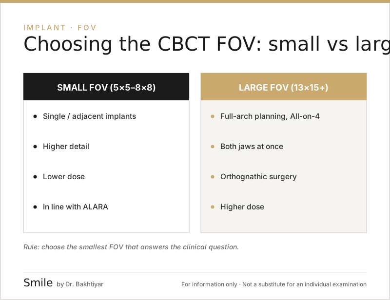
In implant planning, the FOV is chosen according to clinical need: a small FOV for a single implant (usually 4×4–5×5 cm), a medium FOV (8×8 cm) for multiple implants covering one jaw, and a large FOV (10×10 cm and above) for both jaws and full-arch rehabilitation. A small FOV means both a lower dose and higher detail. The position paper of the American Academy of Oral and Maxillofacial Radiology (AAOMR) and the consensus of the European Association for Osseointegration (EAO) recommend cross-sectional imaging of the implant site, but set out limiting the scanned area to the clinical question as the basic rule.
Single implant
For a single tooth gap, a small FOV is the ideal choice: the implant site and the neighbouring structures are fully visible, the dose is minimal, and thanks to the small voxel the nerve canal and adjacent roots are seen more clearly. One condition: boundary structures such as the nerve canal or the sinus floor must fall within the scanned area — the dentist assesses this in advance.
Multiple implants (2–4)
For several implants in one quadrant, an 8×5 or 8×8 cm FOV is usually chosen: the sinus floor and the nerve canal are covered in full.
Implant combined with a sinus lift
For the lateral window approach, the floor, side walls and septa of the maxillary sinus must be visible — an 8×8 cm or larger FOV is needed.
Full arch (All-on-4, All-on-6)
For implants in both jaws, or full rehabilitation such as All-on-4 full-arch implants, a FOV of 10×10 cm or larger is chosen — all the anterior and posterior planning points must be covered.
Orthodontic-surgical preparation
In patients being prepared for orthognathic surgery, the relationship between the two jaws, the jaw joint and the facial bones are assessed — a large (craniofacial) FOV is required.
Practical table
| Clinical situation | Recommended FOV |
|---|---|
| Single implant, small area | 4×4 – 5×5 cm |
| Quadrant (one side) implants | 5×5 – 8×5 cm |
| Sinus lift | 8×8 – 10×10 cm |
| Full arch (All-on-4) | 10×10 cm and above |
| Orthodontic-surgical planning | 15×15 cm or craniofacial |
| TMJ (jaw joint) | 8×8 – 12×9 cm |
| Endodontic problem | 4×4 cm (smallest possible) |
The table is a general guide; the dentist assesses each case individually. For the implant plan itself — bone dimensions, distance to the nerve and sinus, the surgical guide — see: Why are CBCT and digital planning important before an implant?.
Is a CBCT scan brought in from elsewhere usable?
Sometimes yes, sometimes no. The dentist determines the FOV and the operator applies it on the machine. A scan taken elsewhere with a large FOV on the logic of let everything be visible means both an excess dose and, because the detail is lower, it may not be sufficient for planning; and if it does not cover the required area, a repeat scan becomes necessary. The best approach is to take the CBCT with the FOV chosen by the dentist requesting it. In any case, bring the existing scan in DICOM format: the FOV and voxel parameters are recorded in the file metadata, which is important for a second opinion and for planning software (What is DICOM?).
What changes as the FOV gets larger?
- The radiation dose increases — in a 2015 meta-analysis, with standard protocols the mean effective dose for adults was 84 µSv for a small FOV, 177 µSv for a medium FOV and 212 µSv for a large FOV (with wide differences between machines);
- the file size increases (the DICOM volume grows);
- the detail decreases (the voxel gets larger);
- the scan time may lengthen slightly (risk of movement artefacts).
Conclusion
FOV is the key parameter that determines what a CBCT scan will show. The right choice leads both to the right diagnosis and to minimal radiation. The practical rule is simple: choose the smallest area that covers the clinical problem. This is both safer for the patient and more diagnostic for the dentist.
In Baku, a CBCT scan can be taken at our clinic with various FOV options (small, medium, large) and Low Dose protocols.
Book an appointment: Online appointment form
Frequently asked questions (FAQ)
1. Which FOV has the lowest dose?
The smallest FOV (e.g. 4×4 cm). The dose is roughly in the range of 0.02–0.05 mSv.
2. Is a large FOV always better?
No. More anatomy is captured, but the dose also rises. It is chosen only when there is a clinical need.
3. Can the patient choose the FOV?
No. The FOV is chosen by the dentist/radiologist based on the clinical indication.
4. Does a smaller FOV increase accuracy?
As a general rule, a small FOV allows a smaller voxel size, which leads to higher detail — this is particularly important for endodontic and periodontal work.
5. Are FOV and voxel the same thing?
No. FOV is the size of the volume to be scanned (a cylinder); the voxel is the size of the smallest 3D pixel that makes up that volume.
6. Is the smallest FOV always suitable for a single implant?
In most cases yes, but boundary structures such as the nerve canal or the sinus floor must be fully visible. The dentist assesses this before the scan.
7. Will the CBCT scan I bring from elsewhere be usable?
If it covers the required area and the detail is sufficient — yes. If it was taken with a very large FOV and low detail, or the area does not cover the planned site, a repeat scan may be needed.
Author: Dr. Bakhtiyar Aliyev — dentist. This article has been reviewed by a dentist (last updated: 2026-07-06).
Sources
- Position statement of the American Academy of Oral and Maxillofacial Radiology on selection criteria for the use of radiology in dental implantology with emphasis on cone beam computed tomography — American Academy of Oral and Maxillofacial Radiology (OOOO), 2012
- E.A.O. guidelines for the use of diagnostic imaging in implant dentistry 2011. A consensus workshop organized by the European Association for Osseointegration — Clinical Oral Implants Research, 2012
- Effective dose of dental CBCT — a meta analysis of published data and additional data for nine CBCT units — Dentomaxillofacial Radiology, 2015
This article is for general information only and does not replace an individual diagnosis, examination or treatment. Consult a doctor for a decision about your case.