

Why are CBCT and digital planning important before an implant?
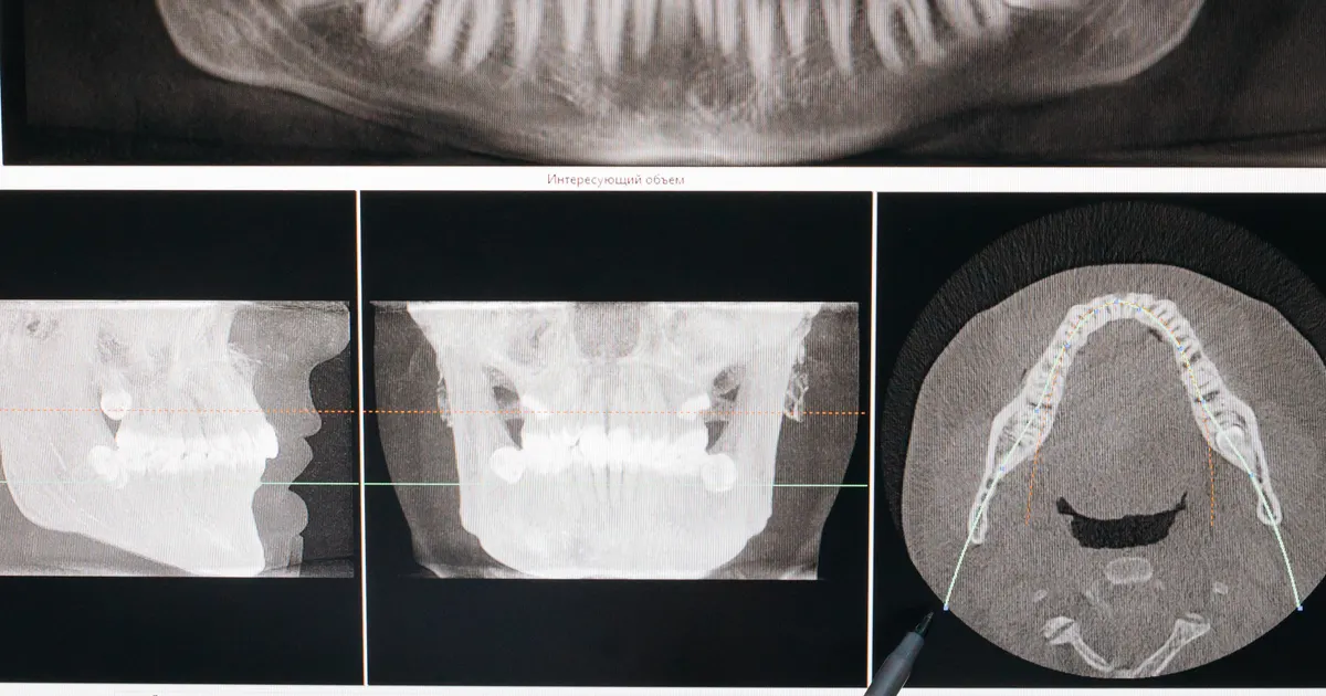
Cone-beam computed tomography (CBCT) shows the jaw in three dimensions: the height and width of the bone, the canal carrying the nerve in the lower jaw, and the condition of the sinus (maxillary sinus) in the upper jaw. When this information is combined with an intraoral scan and the design of the future tooth, the position of the implant is determined before surgery, and a surgical guide is made if needed. This way, surgery rests on a ready plan rather than on unexpected findings. A scan is not prescribed automatically for everyone, but on clinical indication.
How a CBCT scan is performed and how we approach dose is described on the 3D diagnostics service page.
Key points
- A conventional (two-dimensional) X-ray does not show the thickness of the bone or the exact position of structures such as the nerve and the sinus. Cross-sectional 3D imaging fills this gap.
- Digital planning makes it possible to place the implant where the future tooth requires it, rather than simply «wherever there is bone».
- A surgical guide transfers the plan to the mouth. Studies show that with a guided implant the deviation between the plan and the actual position is small, but not zero — which is why a safety margin is maintained.
- International guidelines recommend a scan not for everyone, but in justified cases. The decision is made after an examination and a review of the existing images.
How does CBCT differ from a conventional X-ray?
A panoramic or small (periapical) X-ray shows the jaw flat, in a single plane. It gives an idea of bone height, but the thickness in the lip–tongue direction is not visible. Structures overlap one another and the image comes out somewhat magnified.
CBCT, on the other hand, shows the jaw in slices — cross-sectional, longitudinal and horizontal. At the point where the implant will be placed, the shape of the bone, its concavities and the neighbouring structures are seen separately. We have a separate article on how the machine works and the rules for scanning: what is CBCT (3D imaging). Which images are generally taken before an implant is explained in the next section.
Which X-rays are taken before an implant?
In implant planning each of the three imaging methods has its own place; most often they complement one another.
Panoramic X-ray (OPG) — the initial overview
It shows both jaws, all the teeth, the lower border of the sinuses and the approximate course of the nerve canal in the lower jaw in a single image. It is useful for an initial assessment: teeth that need extraction, impacted teeth, cysts and large areas of inflammation are visible.
When is a panoramic image not enough? The image is two-dimensional — it does not show the width of the bone in the lip–tongue direction or its precise density, structures overlap and dimensions come out somewhat magnified. Wherever the distance to the nerve and the sinus matters, with multiple implants, and when planning a sinus lift and bone grafting, a panoramic X-ray alone is not enough. Only in rare cases — a single-tooth gap with clearly abundant bone, far from neighbouring structures — may the dentist be satisfied with a panoramic X-ray; this decision is made after an examination.
CBCT — the basis of the plan
Cross-sectional 3D imaging makes it possible to measure the height and width of the bone in millimetres, to locate the nerve canal, the sinus floor and the incisive canal precisely, and to see the distance to neighbouring roots. In a sinus lift, the shape of the sinus floor, thickening of the mucous membrane and partitions (septa) are additionally assessed — the choice between a lateral window and a crestal approach is based on this (bone grafting and sinus lifting). The field of view (FOV) is chosen according to clinical need: small for a single implant, medium for multiple implants and a sinus lift, large for a full jaw — in detail: what is FOV and how it is chosen.
Periapical X-ray — supporting
This is a small image showing the root of one or two teeth in detail. It is needed when inflammation is suspected at the root of a tooth to be extracted, or to assess the condition of neighbouring teeth; it is not the main examination for implant planning.
Should the image be taken at the clinic or elsewhere?
Where possible, at the clinic that prescribes it. The dentist chooses the FOV, the scan area and sometimes a radiographic guide (a template showing the position of the future tooth) to suit the clinical purpose. A scan taken elsewhere on a «let us see everything» basis with a large FOV, or one that does not cover the required area, sometimes has to be repeated. If there is a sufficiently recent image covering the required area, a repeat is not needed — be sure to bring the DICOM file.
How is digital planning built?
Digital planning is not simply looking at a scan. Several sets of data are combined in a single virtual model:
- CBCT data (DICOM file): bone volume, the nerve and the sinus, and findings such as old inflammation or a retained root fragment.
- Intraoral scan: the surfaces of the teeth and gums and the way the teeth come together.
- Design of the future tooth: the shape and position of the tooth (digital design or diagnostic model).
These three layers are superimposed and the direction of the implant is chosen according to the future tooth. This is called prosthetically driven planning. Once the plan is ready, either a classic surgical protocol is chosen, or a surgical guide is made on the basis of the plan.
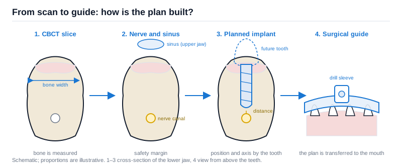 On the slice the bone is measured, the nerve and the sinus are marked, the implant is positioned according to the future tooth, and the guide transfers the plan to the mouth. Proportions are schematic.
Which questions does the plan answer in advance?
Bone volume and quality
The length and width of the implant must be chosen to suit the available bone. On the slice, the width and height of the bone are measured and the ratio between the hard outer layer (cortical bone) and the porous inner part is assessed. This gives an idea of how firm the initial stability of the implant will be. If there is not enough bone, the options — a graft, a sinus lift, a narrower or shorter implant, changing the position — are discussed in the article what options are there when there is not enough bone.
Distance from the nerve and the sinus
In the lower jaw the nerve canal and the mental foramen, and in the upper jaw the sinus and the nasal cavity, define the limits for the implant. On the 3D image these structures are marked in the planning software and a safety margin is maintained between them and the implant. In the lower jaw, proximity to the nerve carries a risk of numbness in the lip — which is why this step is particularly important.
The position and axis of the implant
Even if the implant sits correctly in the bone, an incorrect axis creates difficulties at the prosthetic stage. For example, in a front tooth the screw hole may fall on the visible surface of the tooth, the crown may turn out wider or more angled than it should be, and cleaning may become difficult. At the planning stage, the angle and depth of the implant and its distance from the neighbouring teeth are checked in advance.
Unexpected findings
Sometimes during planning a retained root fragment, an area that has not healed after an earlier extraction, a trace of inflammation within the bone or thickening of the sinus mucous membrane is seen at the implant site. These can change the order of treatment — for instance, the inflammation is resolved first and the implant is placed afterwards.
What does a surgical guide (guided surgery) offer?
A surgical guide is a template that transfers the virtual plan to the mouth; it seats over the teeth, the gums or the bone. The direction and depth of the drills are limited by the channels in the guide.
What to expect:
- Closeness to the plan. A 2024 systematic review and meta-analysis (BMC Oral Health) shows that in computer-assisted placement the mean deviation between the planned and the actual position is at the millimetre level at the entry point and the apex, and a few degrees in angle. Fully guided protocols (all stages with the guide) turn out more accurate than partially guided protocols.
- The deviation is not zero. Precisely for this reason, when the plan is drawn up a reserve margin is maintained between the implant and the nerve and sinus, and the surgeon reassesses the situation during surgery.
- Sometimes less intervention. With suitable anatomy, a protocol without cutting the gum (flapless) becomes possible. However, this is not suitable for everyone; the decision is made individually.
A guide does not replace the surgeon's experience; it is a tool for carrying out a well-constructed plan. What to look for when choosing a clinic and a dentist — we have written about this in the article choosing an implantologist.
Is a scan mandatory for every implant?
Every examination involving radiation must be justified. International guidelines — the imaging consensus of the European Association for Osseointegration (EAO) and the position paper of the American Academy of Oral and Maxillofacial Radiology (AAOMR) — propose the following approach: first the clinical examination and the existing conventional images are assessed; if questions remain and the answer will change the plan, 3D imaging is prescribed.
In practice, a cross-sectional assessment of the implant site is often justified, because there is no other way to measure the width of the bone and the exact position of the nerve/sinus. But this does not mean «a new scan at every visit». If there is a sufficiently recent image covering the required area, a repeat scan is usually not necessary.
What are the limitations of 3D imaging?
A scan is a powerful tool, but it does not show everything and it does not build the plan on its own:
- Artefacts. Metal crowns, posts and existing implants create streaks on the image; that area becomes harder to read.
- Movement. Even a small movement of the head during the scan blurs the image and can cause measurement error.
- Soft tissue is poorly visible. The thickness of the gum and the width of the band of firm (keratinised) gum are assessed by examination and scanning.
- Bone density is approximate. Grey-scale values in CBCT are not standardised as they are in medical computed tomography. The final view on density is formed together with the resistance felt during drilling.
- Radiation. Every scan must be justified, and the scanned area chosen as narrow as possible.
That is why a plan is always a combination of three sources: the clinical examination, the imaging and the goal for the future tooth. Even if everything looks ideal on the image, an implant decision is not considered complete without taking the bite, hygiene habits and general health into account.
How does the plan connect to the prosthetic stage?
The digital plan continues into the prosthetic stage. Based on the intraoral scan, the abutment and the crown are designed and produced with CAD/CAM technology. We have explained this workflow in the article a crown in one day with CAD/CAM. For front teeth the plan is linked to digital smile design — the position of the implant is chosen according to the contour of the future tooth.
In our clinic, the implant plan is built on 3D imaging, an intraoral scanner and digital design. The dental implantation process is agreed with you step by step after the consultation and imaging. For the structure of an implant and the stages involved, you can read the article what is a dental implant.
Frequently asked questions
I have had a panoramic X-ray, do I still need a scan? It depends on the clinical question. A panoramic image shows the general situation. If the width of the bone, the exact position of the nerve or the condition of the sinus raises questions, 3D imaging is prescribed. The dentist makes the decision after an examination.
How long is a scan result valid for? Usually 6–12 months. Because bone changes rapidly in the first months after a tooth extraction, the planning scan is often taken several months after the extraction, close to the time of implant placement.
Is the radiation from a scan dangerous? The dose of a dental CBCT depends on the FOV and is many times lower than that of medical computed tomography; when it is taken on indication and with the smallest possible field, the risk is statistically negligible. In detail: is CBCT radiation high?.
Is one scan enough for several implant sites? If the planned sites fall within the limits covered by the scan, one image is usually sufficient. If the situation changes or a long time passes, a repeat assessment may be needed.
Can an implant be placed without a guide? Yes. A guide is not compulsory; in most cases a freehand protocol based on the plan is used. A guide is particularly useful close to the nerve/sinus, in front teeth and with multiple implants.
Does a digital plan guarantee the result? No. Planning is a means of increasing predictability. The result is also affected by healing, hygiene, general illnesses, smoking and the load from the bite.
Can I read the scan myself? Reading the slices takes experience. What matters for you is that the dentist explains the plan in plain language: how many implants, in which positions, whether an additional procedure is needed and in what order the stages will go.
Conclusion
In implant treatment the main work is done before surgery. 3D imaging shows the bone, the nerve and the sinus; the digital plan determines the position of the implant according to the future tooth; the surgical guide transfers the plan to the mouth. These steps do not turn the result into a guarantee, but they base decisions on measurable information rather than on guesswork. To assess your individual situation, your existing images and your general health information are reviewed together during the consultation.
Sources
- E.A.O. guidelines for the use of diagnostic imaging in implant dentistry 2011. A consensus workshop organized by the European Association for Osseointegration — Clinical Oral Implants Research, 2012
- Position statement of the American Academy of Oral and Maxillofacial Radiology on selection criteria for the use of radiology in dental implantology with emphasis on cone beam computed tomography — American Academy of Oral and Maxillofacial Radiology (OOOO), 2012
- Accuracy of implant placement with computer-aided static, dynamic, and robot-assisted surgery: a systematic review and meta-analysis of clinical trials — BMC Oral Health, 2024
This article is for general information only and does not replace an individual diagnosis, examination or treatment. Consult a doctor for a decision about your case.