

What can be done if there is not enough bone for an implant?
Having little bone often does not mean that an implant is impossible. Based on an examination and 3D imaging, several routes are considered: bone grafting, a sinus lift in the upper back jaw, a short or narrow implant that suits the available bone, tilted placement, and, in a fully edentulous jaw, All-on-4 type concepts. Which one suits you depends on the size and location of the deficiency, the quality of the bone and the planned prosthesis. In some severe cases the options may be limited.
Key points
- Not enough bone is a relative rather than an absolute concept: bone height, width and quality are assessed together with the size and position of the planned implant.
- The solution is chosen according to the extent of the deficiency: either we adapt to the available bone (a short/narrow implant, tilted placement), or we augment the bone.
- Bone grafting is sometimes carried out at the same appointment as the implant and sometimes as a separate stage — in the latter case treatment takes longer.
- An ordinary X-ray is not enough for this decision: 3D imaging shows the position of the sinus and the lower jaw nerve, and the real bone volume.
- Smoking, uncontrolled diabetes and past periodontitis can affect healing and the long-term outcome, so they are built into the plan.
What does not enough bone actually mean?
An implant is a titanium post placed inside the jawbone, and there must be healthy bone of a certain thickness around it. That is why the dentist looks at three things:
- Height — the vertical distance available for the length of the implant. This means the distance up to the sinus cavity above and to the canal of the lower jaw nerve below.
- Width (thickness) — in addition to the diameter of the implant, a reserve for the bone wall covering it on the cheek side.
- Quality and position — the density of the bone and where the missing part is located. Even if there is enough bone overall, there may be too little at the exact point the prosthesis requires.
In other words, the not enough decision is not made in a vacuum; it is made in relation to the planned tooth and prosthesis. In an ITI consensus report the thickness of the bone wall surrounding the implant is considered important for outcomes; if that wall is thin or missing, guided bone regeneration (GBR — restoring bone by covering the material with a membrane) can be helpful.
Why does bone shrink?
The most common reason is simple: after a tooth is extracted, the bone in that area gradually shrinks, and the longer the area stays without a tooth, the greater the loss. There are other reasons too: gum disease (periodontitis) lasting many years, trauma, wearing a removable denture for years, and downward expansion of the sinus cavity in the upper jaw (pneumatisation).
How the decision is made: plan first, surgery second
In the modern approach the sequence is as follows: first the planned prosthesis is decided, then the ideal position of the implant is determined, and only after that is it assessed whether there is enough bone in that position. This is why 3D imaging and digital planning before an implant is a key step — it is exactly what shows the real extent of the deficiency and which solution is possible.
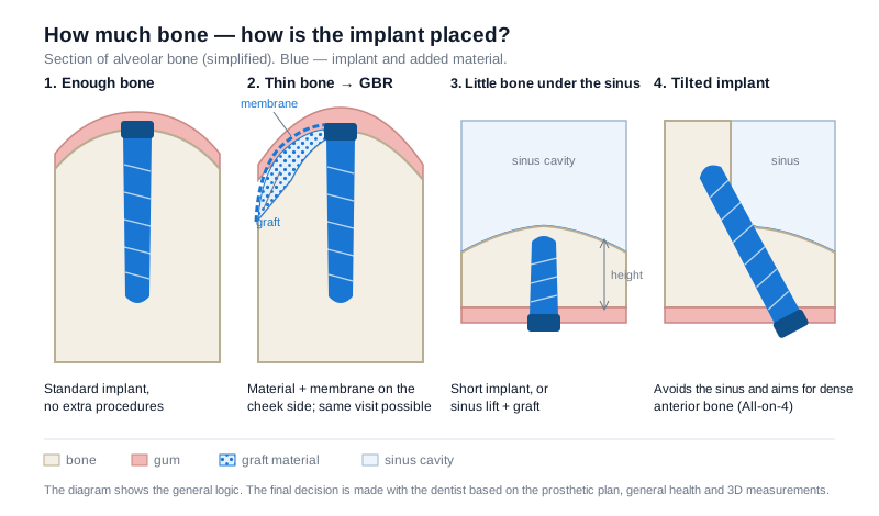
Typical scenarios in a bone cross-section and the solution discussed in each. The diagram is simplified; an individual plan is built through an examination.
Option 1 — Bone grafting (graft and GBR)
Bone material is added to the deficient area and is usually covered with a membrane (a thin film). Over time the material is replaced by your own bone, or fuses with it to create a foundation for the implant. The material can come from your own bone, from a donor, from an animal source, or be synthetic; the choice is made according to the size and shape of the defect.
With small defects, grafting may be possible at the same appointment as the implant. With larger defects a staged plan is chosen: first the bone is augmented, and once it has healed the implant is placed. Patient information from NHS hospitals states that, before moving from the graft to the implant stage, several months (roughly 3–6 months) are usually allowed for the bone to heal. A detailed explanation of materials, techniques and healing is in the bone grafting article.
Option 2 — Sinus lift in the upper back jaw
In the back part of the upper jaw, the main factor limiting height for an implant is the sinus cavity. In this case the sinus membrane is carefully lifted and bone material is placed in the space created beneath it. The procedure can be carried out from the cheek side (lateral) or directly through the implant socket (crestal). The stages, healing and possible risks are explained separately in the sinus lift article.
Option 3 — Short and narrow implants
Sometimes, instead of augmenting the bone, it is possible to choose an implant sized to suit the available bone. According to a systematic review and meta-analysis published in 2022, in selected cases in the posterior region of the upper jaw short implants can be an alternative to standard-length implants placed together with a sinus lift: over the follow-up periods compared, no clear difference was found in implant survival or bone loss around the implant. The authors also note that longer observations are needed.
The practical advantage of this approach is a shorter surgical stage and shorter healing. Its limitation is that a short implant is not suitable everywhere: bone quality, the function of the tooth, the bite load, the height of the planned crown and the number of implants must all be taken into account. Narrow implants are considered along the same lines — in a thin bone ridge — but as the diameter decreases, the question of mechanical load becomes more important.
Option 4 — Tilted implants and the All-on-4 concept
Bone does not shrink to the same degree everywhere: usually more bone remains in the front region and away from the sinus. Tilted placement makes use of exactly this — the implant is angled away from the sinus or the nerve and directed into a denser part of the available bone. In a fully edentulous jaw, the All-on-4 approach built on this principle allows some patients to avoid extensive grafting. However, it is not a universal solution: the number of implants, the type of prosthesis and the distribution of load are planned individually.
Option 5 — Special surgical solutions in severe atrophy
When bone loss in the upper jaw is very severe, zygomatic implants, which take their support from the cheekbone (zygoma), may be discussed. A review published in 2021 (an overview of systematic reviews) presents these implants as a treatment option for the severely atrophic upper jaw, while also reporting that complications such as sinusitis have been recorded. This is a procedure that requires special preparation, experience and facilities, and it is not for every patient. In such cases, referral to a suitable centre is normal practice.
Same appointment, or staged?
This is the question that concerns patients most, because it is what determines the overall duration. The logic is this: if the implant is firmly stable in the bone at the moment it is placed (this is called primary stability), then in small and moderate defects grafting and the implant can be carried out at the same appointment. If such stability is not expected, or the defect is large, a staged plan is chosen. With a staged plan treatment takes longer, but in return the bone foundation is prepared in a more predictable way. During this period, especially in the front tooth region, a temporary solution can be planned.
Factors that can affect the outcome
When the plan is drawn up, not only anatomy but also factors affecting healing are taken into account: smoking, uncontrolled diabetes, active gum disease, medicines affecting bone metabolism (for example, bisphosphonates), night-time clenching and hygiene. Some of these can be managed before treatment, and this improves the chances of the plan succeeding. When a dental implantation plan is being prepared, it is important to discuss these matters openly from the outset.
Frequently asked questions
A dentist told me there is no bone and an implant is not possible — is it worth getting a second opinion? Yes, this is a normal step. Assessment methods can differ: a conclusion drawn from an ordinary X-ray may change after 3D imaging. When you go for a second opinion, take your existing images with you.
Does the graft have to be taken from my own bone? No. Your own bone is the preferred option in some cases, but donor, animal-derived and synthetic materials are also widely used. The choice depends on the size and shape of the defect and on the planned technique.
Are there cases where an implant is placed without bone grafting? Yes. If the deficiency is small, a narrow or short implant and, in suitable cases, tilted placement can reduce the need for grafting. Whether this is possible is shown by 3D measurements and the prosthetic plan together.
I have worn a removable denture for many years and the bone has shrunk a lot — what options are left? In such cases, what is usually discussed is either large-volume grafting, or concepts that make maximum use of the available bone (tilted implants, full-arch solutions). Sometimes a removable denture seated on implants is the more realistic choice.
How much does bone grafting extend treatment? It depends on the volume of the graft and on whether it is carried out at the same appointment as the implant. In staged plans, several months are allowed for the bone to heal; the exact duration is shown by follow-up examinations and imaging.
Can the area shrink again after bone grafting? An augmented area is living tissue too: load, hygiene and inflammation all affect it. That is why regular check-ups and professional cleaning remain part of the plan after the implant as well.
Conclusion
Bone deficiency is a situation that comes up often in implantology and can usually be managed. The choice is built along two lines: augmenting the bone, or choosing a concept that suits the available bone. Which is more appropriate is determined by anatomy, the prosthetic plan and your own health factors together. For an assessment of your individual situation, a consultation and 3D imaging are the best starting point.
Sources
- Group 1 ITI Consensus Report: The role of bone dimensions and soft tissue augmentation procedures on the stability of clinical, radiographic, and patient-reported outcomes of implant treatment — Clinical Oral Implants Research (ITI Consensus Report), 2023
- Simultaneous placement of short implants versus standard length implants after sinus floor elevation in atrophic posterior maxillae: a systematic review and meta-analysis — International Journal of Implant Dentistry, 2022
- Zygomatic implants placed in atrophic maxilla: an overview of current systematic reviews and meta-analysis — Maxillofacial Plastic and Reconstructive Surgery, 2021
- Bone grafting for dental implants — Guy's and St Thomas' NHS Foundation Trust
This article is for general information only and does not replace an individual diagnosis, examination or treatment. Consult a doctor for a decision about your case.