

What is bone grafting and when is it needed before an implant?
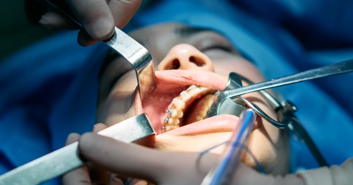
Bone grafting (augmentation) means placing a special material into the deficient area when the jawbone is not sufficient for an implant. This material acts as a scaffold for new bone to form and is often covered with a membrane (a thin barrier). The procedure can be carried out at the same session as the implant or separately, beforehand. Whether you need it is shown by a clinical examination and 3D imaging (CBCT); the decision is individual for every patient.
Key points
- Bone grafting is not a single «type of operation» — it is a set of techniques that vary according to the type of defect.
- Materials fall into four main groups: autogenous (your own bone), allogeneic (donor), xenogeneic (animal-derived) and synthetic.
- According to ITI consensus documents, horizontal augmentation is more predictable; vertical augmentation is more complex and carries a higher likelihood of complications.
- If height is lacking in the posterior region of the upper jaw, a separate technique — a sinus lift — is used.
- Healing time depends on the site, the size of the defect and the material; rushing does not improve the outcome.
Why does the jawbone shrink?
The alveolar bone — the bone surrounding the roots of the teeth — is maintained thanks to the load placed on the tooth. When a tooth is extracted this load is lost and the bone reduces both in width and in height, particularly during the first months: the area becomes narrower and lower. In a site that has been without teeth for many years, this change is more noticeable.
There are other causes too: severe periodontitis (inflammation of the tissues around the tooth), a long-standing infection at the root tip, trauma, a cyst, expansion of the sinus cavity. The result is the same in all of them — there is less bone than is needed for the implant to be reliably supported. For the overall picture and alternative solutions, you can read the article on options when there is not enough bone for an implant.
What does bone grafting mean?
During bone grafting, bone material is placed into the deficient area. This material is not itself «new bone». Its job is to maintain the space and create a scaffold into which your body's own cells can grow. As the months pass, the material is gradually replaced by your own bone, or it remains within the new bone and integrates with it.
The most commonly used technique for controlling this process is called guided bone regeneration (GBR): the material is covered with a membrane — a thin biological or synthetic barrier. The membrane prevents the faster-growing soft tissue from filling the site and buys time for the bone cells. In the ITI consensus recommendations, placing a membrane is listed as indicated when particulate (granule) material is used.
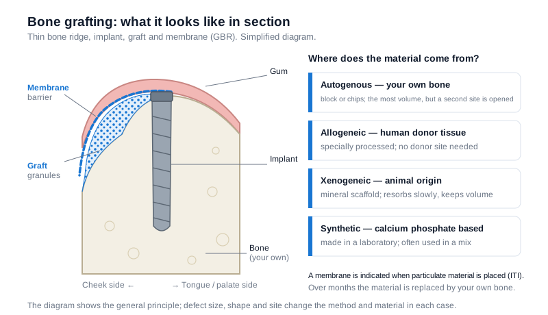 Diagram: an implant in a thin bony ridge, graft placed towards the cheek side and the membrane covering it (GBR). On the right, the four main sources of material.
What types of material are there?
- Autogenous (your own bone). Usually taken from another area of the jaw as a block or as chips. ITI documents report that in horizontal and vertical augmentation, autogenous block grafts provide more volume gain than particulate materials; in return, a second surgical site (the donor site) has to be opened.
- Allogeneic. Donor tissue of human origin that has undergone special processing. It allows volume to be added without opening a donor site.
- Xenogeneic. A mineral scaffold of animal origin; because it resorbs slowly, it helps to maintain volume over a long period.
- Synthetic (alloplastic). Calcium phosphate based materials produced in a laboratory; they require no donor tissue.
In practice these groups are often combined — for example, your own bone chips are mixed with xenogeneic granules. According to the ITI consensus, none of the techniques is unequivocally superior to another in terms of implant survival; the choice is made according to the shape of the defect, the volume required and the anatomy of the site.
When is it needed before an implant?
The need depends on the type of defect. The most commonly encountered situations are the following:
Small defects (dehiscence and fenestration). When the implant is placed, a small part of its surface is not covered by bone. In such cases the material and membrane are placed during the same operation as the implant. The ITI consensus reports that covering this type of defect is effective in reducing the exposed implant surface, but that complete regeneration cannot always be predicted.
Horizontal deficiency. There is enough height, but not enough width to surround the implant. This is the most common scenario and the prognosis is relatively good.
Vertical deficiency. The height of the bone is insufficient — for example, after a long period without teeth or after severe inflammation. ITI documents indicate that the prognosis for vertical augmentation is lower than for horizontal augmentation, while the complication rate is higher. Such cases require separate planning and often a staged approach.
The posterior region of the upper jaw. Here the maxillary sinus (the air cavity beside the nose) limits the height, and a separate technique — a sinus lift — is used. Its stages, the difference between the lateral and crestal approaches and the risks are a separate topic: what is a sinus lift.
Socket preservation at the time of extraction
Placing material into the empty socket immediately after a tooth is extracted is called «socket preservation» (ridge preservation); the aim is to reduce subsequent bone loss. The technique can be useful, but the evidence is not clear-cut. A review published in the British Dental Journal in 2022, citing systematic reviews, reports that in the months after extraction (up to the implant stage) the difference measured in ridge width between socket preservation and simple extraction is in the region of one millimetre, and no clear advantage has been shown in terms of the need for additional grafting when the implant is placed. For that reason the decision is made case by case: whether the tooth is in the anterior or posterior region, the integrity of the socket walls and the future plan are all taken into account. Whether extraction and implant placement are possible on the same day is a separate topic: same-day implant after extraction.
Same stage, or separately?
There are two scenarios and both can be correct:
- Same stage (simultaneous): if the existing bone can hold the implant with initial stability, the graft and the implant are carried out in a single operation. The overall treatment time is shorter.
- Separately (staged): if the defect is large or the primary stability of the implant cannot be ensured, the bone is augmented first, healing is awaited, and the implant is placed afterwards.
The choice is made by measurement, not by eye. The width and height of the bone and the position of the sinus and of the lower jaw nerve are assessed with cone-beam computed tomography (CBCT) — more on this here: CBCT and digital planning before an implant. The surgical stage is planned as part of dental surgery.
How does healing go?
The operation is usually carried out under local anaesthesia; if necessary, sedation can be discussed. In the first few days, swelling, tenderness in the area and sometimes bruising are to be expected and pass with time.
General rules for the following days:
- Eat soft food during the first few days and keep the surgical site free of load.
- Follow the medication and mouth rinse regimen prescribed by your dentist.
- Do not poke the area with your tongue or a brush; protect the sutures.
- Avoid smoking — smoking is one of the factors that adversely affect wound healing and the acceptance of the graft.
- Attend your follow-up appointments on time.
The time needed for bone to form is not the same for everyone: it is relatively short for small defects and longer for extensive or vertical augmentation. Whether it is ready is checked with an examination and repeat imaging.
Risks and limitations
Bone grafting is a surgical procedure, and no surgical procedure comes with a guaranteed outcome. Possible complications include: opening of the suture line and exposure of part of the material, infection, less volume gain than expected and, rarely, failure of the graft to be accepted. The ITI consensus notes that as the defect becomes more complex, the complication rate and the need for additional grafting increase.
Sometimes declining augmentation altogether is also the right plan: short or narrow implants, angled placement, or a different prosthetic solution can give the same functional result with less intervention. These options are discussed at the planning stage together with the overall aim of dental implantation treatment.
Frequently asked questions
When can an implant be placed after bone grafting? There is no single time frame — the size of the defect, the material and the site are decisive. In small defects the implant is often placed during the same operation as the graft; in extensive augmentation a separate period is allowed for healing and readiness is confirmed with a repeat examination.
Does it have to be taken from my own bone? No. Autogenous bone can be the preferred choice in some defects, particularly when volume is needed in block form. In many cases, however, allogeneic, xenogeneic or synthetic materials, or a mixture of them, are sufficient.
Can the graft material be rejected by the body? Dental graft materials are not living tissue that triggers an immune response, but a processed mineral scaffold; they are not «rejected» in the classic sense. However, integration may fail because of infection, opening of the suture or the material not remaining stable in place — in such cases a repeat procedure may be needed.
Do diabetes or smoking prevent bone grafting? They do not necessarily prevent it, but they change the risk. Uncontrolled diabetes and smoking are among the factors that affect wound healing; before planning, discuss your general health and the medicines you take (particularly medicines that affect bone metabolism) with your dentist.
Are bone grafting and a sinus lift the same thing? No. A sinus lift is a specific technique relating to the posterior region of the upper jaw: the sinus membrane is lifted and material is placed underneath it. Bone grafting is a broader concept and can relate to horizontal or vertical deficiency anywhere in the jaw.
Conclusion
Bone grafting is not an «add-on» to an implant but part of the plan: the aim is not simply to place the implant, but to place it in the correct position and surrounded by sufficient bone. The type of defect determines the technique, and the technique determines the timetable. To assess your individual situation, a consultation based on a clinical examination and 3D imaging is advisable.
Sources
- Bone Augmentation Procedures in Localized Alveolar Ridge Defects — Consensus Statements — ITI (International Team for Implantology) Consensus Conference
- Is there clinical evidence to support alveolar ridge preservation over extraction alone? A review of recent literature and case reports of late graft failure — British Dental Journal, 2022
This article is for general information only and does not replace an individual diagnosis, examination or treatment. Consult a doctor for a decision about your case.