

A sinus lift is a procedure in which the thin membrane covering the floor of the sinus (the maxillary sinus) in the side region of the upper jaw is raised, and bone material is placed in the space created beneath it. The aim is to restore the bone height needed for an implant. There are two main techniques: lateral (through a small window on the cheek side) and crestal (through the tooth site, via the implant socket). Which one is chosen depends on the height of the remaining bone, the anatomy of the sinus and the size of the planned implant.
Key points
- A sinus lift is performed only in the back (side) part of the upper jaw — in the area of the premolars and molars; there is no such procedure in the lower jaw.
- If the remaining bone is relatively good, the less invasive crestal technique is chosen; if the bone has decreased considerably, the lateral window technique is used.
- Sometimes the implant is placed in the same session as the sinus lift, and sometimes as a separate stage after the bone has matured.
- The most common complication during the procedure is tearing of the sinus membrane (the Schneiderian membrane); in most cases it is managed during the operation itself.
- 3D imaging — CBCT — is essential for planning: residual bone height, sinus septa, wall thickness, the vessel canal and the condition of the sinus are measured; if there is active sinusitis, the operation is postponed.
Why is there not enough bone in the upper jaw?
Above the roots of the side teeth of the upper jaw lies an air-filled cavity — the maxillary sinus. After a tooth is lost, two processes take place at the same time: the alveolar bone that held the tooth gradually resorbs, while the sinus expands downwards (this is called pneumatisation). As a result, the layer of bone remaining between the gum and the sinus floor becomes too thin to hold an implant safely.
A Cochrane systematic review also notes that insufficient bone volume in the back part of the upper jaw is one of the most common obstacles to implant-supported restoration, and that lifting the sinus floor is one of the techniques used to increase this volume. In other words, the root of the problem is not only the anatomy of the sinus, but also the bone resorption that begins after tooth loss.
How is a sinus lift performed?
The procedure is carried out under local anaesthesia, with sedation added if needed. The general principle is the same in both techniques: the thin membrane lining the inside of the sinus is raised without being damaged, and bone material (your own bone, an animal-derived or synthetic graft, most often a mixture of these) is placed in the space beneath it. Over time the material is replaced by your own bone and creates support for the implant. You can read about the types of material in a separate article on bone grafting.
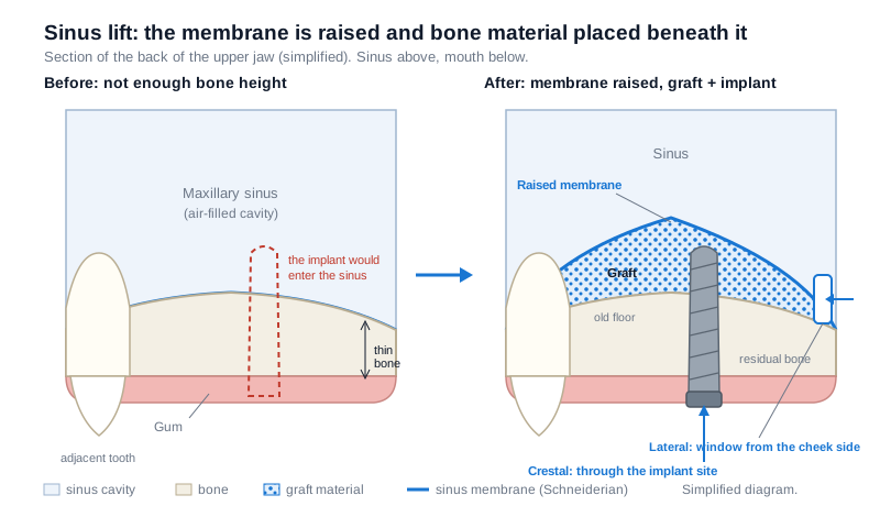 Diagram: on the left, the thin bone remaining under the sinus after tooth loss; on the right, the membrane raised, the graft placed beneath it and the implant positioned. The arrows show the two access routes.
The lateral (side window) technique
The gum is lifted, a small window is opened in the bone wall on the cheek side of the jaw, the membrane is carefully detached and raised, the space is filled with graft and the area is closed. This technique makes it possible to gain more height, which is why it is chosen when the bone has decreased significantly.
The crestal (transcrestal) technique
The sinus floor is pushed upwards with special instruments (osteotomes or drill systems) through the prepared implant socket, without opening a separate window. According to ITI Academy materials, this approach usually requires a certain minimum residual bone height — around 6 mm — and a relatively flat sinus floor. The technique is less traumatic, but the height gained is limited and the surgeon does not see the membrane directly.
| Criterion | Lateral (side window) | Crestal (transcrestal) |
|---|---|---|
| Access route | The bone wall on the cheek side of the jaw | The implant socket prepared at the tooth site |
| Typical indication | When residual bone height is significantly reduced | When residual bone is relatively sufficient (approximately 6 mm and above) |
| Height gained | More | Limited |
| Visibility of the membrane | Direct vision is possible | Vision is limited; worked by feel and X-ray control |
| Placing the implant in the same session | Possible if the residual bone allows primary stability | Most often carried out in the same session as the implant |
| Swelling and recovery | Usually more noticeable | Usually milder |
Who is it suitable for, and who is it not?
A sinus lift is for patients who have lost the side teeth of the upper jaw and want a fixed implant-supported restoration in this area. The decision is based not only on the X-ray image, but also on general health.
Situations that require postponing the procedure or choosing another route:
- acute sinusitis or untreated chronic sinus disease, blockage of the natural drainage pathway of the sinus, polyps and other pathologies — in such cases an ENT specialist's opinion is needed first;
- uncontrolled systemic conditions, a history of radiotherapy to the head and neck area, taking certain medicines that affect bone resorption;
- poor hygiene and untreated gum inflammation — gum treatment and professional cleaning are planned first;
- active smoking is a risk factor in terms of healing and outcome, and is discussed separately at the consultation.
An individual decision is made only after an examination and 3D diagnostics. All scenarios of insufficient bone and the possible alternatives are compared in the article not enough bone for an implant.
The role of CBCT in planning
A sinus lift cannot be planned with an ordinary X-ray: a panoramic image does not show the width of the bone, the condition of the membrane, the septa or the vessel canal. A three-dimensional image answers the following questions before the operation:
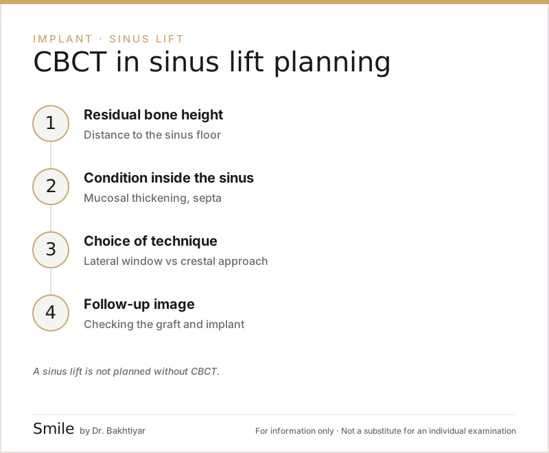
- Residual bone height. The distance from the sinus floor to the alveolar surface is measured in millimetres — this figure determines the choice of technique (crestal or lateral) and whether the implant can be placed in the same session.
- Bone width. If the alveolar ridge is narrow, horizontal bone grafting may also be needed for implant stability, even if the sinus lift is successful.
- The sinus membrane (Schneiderian membrane). Normally it is very thin; thickening (usually more than 2 mm) points to past or active inflammation and increases the risk of tearing — in such a case the operation may be postponed and the sinus treated first.
- Sinus septa and the thickness of the side wall. According to a 2024 systematic review and meta-analysis, the presence of septa is associated with a higher likelihood of membrane tearing; the position of the window is chosen accordingly.
- The vessel canal. The position of the posterior superior alveolar artery running through the side wall is important for the design of the lateral window — if it is cut, heavy bleeding can occur.
- Existing sinus pathology. Mucous retention cysts, polyps, an air-fluid level (active sinusitis), inflammation of dental origin — active sinusitis is a reason to postpone the operation and an ENT specialist's opinion may be required. What can be seen in the sinus is covered in a separate article: can CBCT show sinus problems?.
The scanned area (FOV) is usually chosen at a medium size — the floor and side walls of the sinus must be visible together with the upper jaw; a small FOV is often not enough (what is FOV and how is it chosen). The image is uploaded into the planning software, measurements are taken on the slices, the implant is placed virtually and, if needed, a surgical guide is produced. If the operation was difficult or the membrane was torn, a repeat CBCT may be taken for control before the implant is placed. The general logic of implant planning is described in the article CBCT before an implant.
Can the implant be placed at the same time?
This depends on whether the residual bone is able to hold the implant stable at the initial stage:
- The same session (a one-stage approach): if the residual bone provides primary stability for the implant, the sinus lift and the implant are carried out together. The overall treatment time is shorter.
- A two-stage approach: if there is very little bone, only the graft is placed first; once the material has matured, the implant is placed in a separate session.
In both cases the permanent restoration is not fitted onto the implant straight away — time is needed for osseointegration, that is, for the implant to bond with the bone. To learn about how an implant works, see the article what is a dental implant.
What is healing like?
In the first few days, swelling, slight bruising on the cheek and a small amount of blood-stained discharge from the nose are possible — these are expected reactions. The dentist usually advises the following:
- do not blow your nose forcefully, and keep your mouth open when you sneeze;
- for a while, do not drink through a straw, do not play wind instruments, and avoid diving and heavy physical exertion;
- take the prescribed medicines as directed, and keep the area gently clean;
- avoid smoking.
Maturation of the graft is measured in months rather than days, and depends on the material, the height gained and your own healing characteristics. The exact time is determined by your dentist using follow-up X-rays.
Risks and complications
The most talked-about complication is tearing of the sinus membrane. When it is detected during the operation, it is usually managed with membrane coverings and a change of technique; in some cases the procedure is postponed. Other possible problems include: infection and a flare-up of sinusitis, loss of part of the graft, prolonged swelling, and, rarely, graft material passing into the sinus cavity.
Most of these risks are reduced by careful case selection, precise 3D planning and following the post-operative instructions. The likelihood of complications is individual and should be explained at the consultation in terms of your own anatomy. In oral surgery treatments, discussing the risks in writing before the operation is the standard approach.
Are there alternatives to a sinus lift?
Not every bone deficiency requires a sinus lift. Depending on the situation, the following approaches can be discussed:
- Short implants: if the residual bone height is above a certain threshold, it may be possible to choose a shorter implant without a sinus lift. The Cochrane review stresses that research in this field is still limited and that the results should be interpreted with caution.
- Implants placed at an angle: using the available bone by passing the implant to the side of the sinus.
- Support points outside the sinus: in a completely edentulous jaw, in concepts such as All-on-4, the back implants are placed at a tilt.
The choice depends not only on the amount of bone, but also on the number of teeth, the bite and the planned restoration.
Frequently asked questions
Does the risk of sinusitis increase after a sinus lift? In patients with a healthy sinus who follow the instructions, this is not an expected outcome. However, if there is pre-existing sinus disease, the risk increases — which is why the condition of the sinus is assessed at the planning stage and, where necessary, we work together with an ENT specialist.
Is pain felt during the operation? The procedure is carried out under local anaesthesia, so pain is not expected during the operation; a sensation of pressure and movement may be felt. In the following days, discomfort usually passes with the prescribed medicines.
Is the crestal technique less effective than the lateral one? No, their indications are simply different. The crestal approach is less invasive, but gains limited height and requires a certain amount of residual bone; if there is very little bone, the lateral technique is more predictable.
Should my own bone be used for a sinus lift? Not necessarily. According to systematic reviews, bone substitutes can in many cases be used as an alternative to your own bone; the choice is made according to the size of the area and the planned stages.
How long does an implant placed with a sinus lift last? Its lifespan depends not on the fact of the sinus lift, but on hygiene, follow-up visits, smoking and general health. Regular check-ups and professional cleaning are needed to maintain the long-term result.
Conclusion
A sinus lift is a supporting surgical procedure that makes an implant possible by restoring bone height lost in the back part of the upper jaw. The choice between the lateral and crestal techniques is based on the residual bone and the anatomy of the sinus, while whether the implant is placed in the same session is based on primary stability. Correct diagnostics and a staged plan determine the outlook for this procedure. A consultation and 3D imaging are the first step in assessing your individual situation.
Sources
- Interventions for replacing missing teeth: augmentation procedures of the maxillary sinus — Cochrane Database of Systematic Reviews, 2014
- Association between sinus septa and lateral wall thickness with risk of perforation during maxillary sinus lift surgery: A systematic review and meta-analysis — PLOS ONE, 2024
- Transcrestal Sinus Floor Elevation — ITI Academy Learning Module — International Team for Implantology (ITI) Academy
This article is for general information only and does not replace an individual diagnosis, examination or treatment. Consult a doctor for a decision about your case.