

Yes. CBCT provides a 3D image of the maxillary sinus in the upper jaw and clearly shows thickening of the Schneiderian membrane, fluid, polyps, cysts and the condition of the bony walls. In cases of dental (odontogenic) sinusitis in particular, CBCT is the first choice — because it assesses both the teeth and the sinus in the same image. For the frontal, ethmoid and other facial sinuses, a conventional head CT is usually superior.
In this article we explain the role of CBCT in sinus diagnosis, which problems it can reveal and when further assessment by an ENT specialist is needed.
The maxillary sinus and tooth roots — the anatomical link
The roots of the upper back teeth (premolars and molars) lie very close to an air-filled space called the maxillary sinus. In some patients the tooth root projects into the sinus (with only a thin layer of bone, or with no bone at all). This anatomical proximity has two important clinical consequences:
- Inflammation from a tooth can spread into the sinus → odontogenic sinusitis.
- Sinus surgery or trauma can affect the teeth.
CBCT shows this relationship clearly — it is possible to measure the millimetre-level distance between each tooth root and the sinus wall.
Indicators of sinus condition on CBCT
1. Thickness of the Schneiderian (sinus) membrane
The inner lining of the sinus is called the Schneiderian membrane. Normally it is very thin (less than 1 mm). On CBCT:
- ≤2 mm — normal / minimal finding.
- 2–5 mm — local irritation, a previous sinus problem.
- >5 mm — a significant pathological sign (chronic inflammation, allergy, odontogenic source).
- Completely filled with fluid — acute sinusitis.
2. Air-fluid level
Fluid collects inside the sinus and a level appears depending on the position of the head. This is the classic sign of acute bacterial sinusitis.
3. Polyps and pseudocysts
- Mucous retention pseudocyst — a soft, dome-shaped formation rising from the sinus floor, usually symptomless.
- Polyp — more diffuse, multiple or obstructive.
4. Changes in the bone structure
- Thinning of the sinus wall or a defect.
- Cortical hardening (sclerosis) — a sign of chronic inflammation.
- Sinus septa (dividing walls) — important for planning implants and sinus lifting.
Odontogenic sinusitis — the ideal indication for CBCT
Odontogenic sinusitis is inflammation spreading into the sinus from the upper teeth. The causes are:
- Inflammation at the tooth root (periapical abscess)
- Root canal material passing into the sinus
- Entry into the sinus during implant placement
- An oro-antral communication at the site of an extracted tooth
- Spread of a tooth root cyst into the sinus
Because CBCT shows both in the same image, it is invaluable in diagnosing odontogenic sinusitis. An international multidisciplinary consensus document (2021) regards imaging that shows the dental source and the sinus together — CBCT or CT — as essential for diagnosing odontogenic sinusitis, alongside the clinical signs; in one-sided sinusitis that does not respond fully to antibiotics, a dental cause should be considered especially carefully, since it is often treated at first as ‘ordinary sinusitis’.
When is CBCT recommended for the sinuses?
- One-sided sinus complaints (headache, pressure under the eye, blocked nose).
- Sinus signs together with dental inflammation in the upper jaw.
- Implant planning (see Why CBCT is essential for dental implants).
- Sinus lifting (raising the sinus floor) planning.
- Pain spreading to the sinuses after root canal treatment.
- Suspected oro-antral communication.
- Unexplained tooth pain of sinus origin.
Plain sinus X-ray, CBCT and head CT — which is for what?
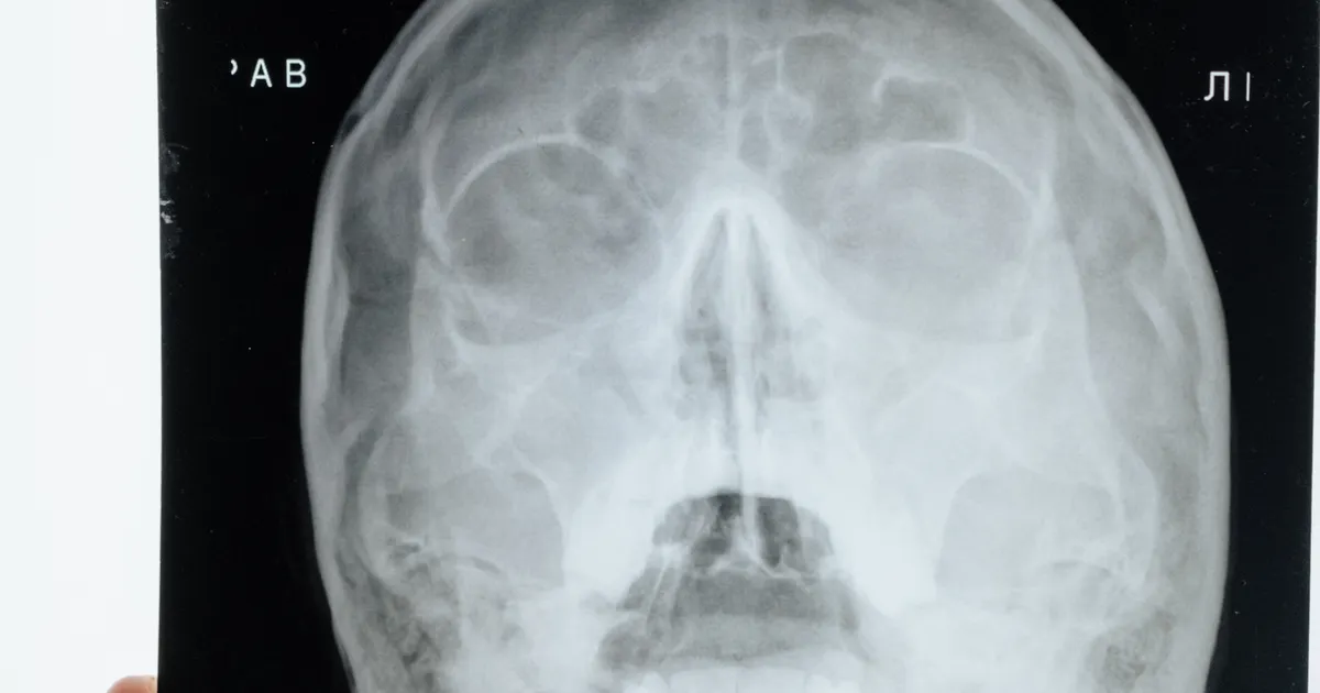
A plain sinus X-ray (the ‘Waters’ and ‘Caldwell’ positions in ENT practice) is a classic two-dimensional X-ray: it can show a fluid level in acute sinusitis, how well the sinus is filled with air, large polyps and cysts, and serious fractures or deformities of the walls. It is quick and low-dose (~20–40 µSv), but it does not show the relationship between the teeth and the sinus, the thickness of the mucous lining or bone detail — so it is not sufficient for questions about odontogenic sinusitis, oro-antral communication, implants and sinus lifts.

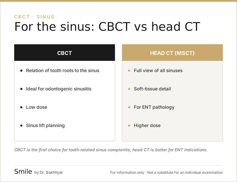
CBCT shows the sinus in three planes (axial, sagittal, coronal): the distance between each tooth root and the sinus floor is measured in millimetres, and membrane thickness, pseudocysts, septa and involvement of the ostiomeatal complex (the drainage pathway of the sinus) are assessed. The dose depends on the FOV — in a 2015 meta-analysis, the average dose for a medium FOV with a standard protocol was ~177 µSv, and it is lower with small FOV and Low Dose modes.
A conventional head CT (MSCT) is the ENT specialist’s standard method for all the facial sinuses (frontal, ethmoid, sphenoid), vascular problems and extensive inflammatory processes; its dose is many times higher than that of CBCT.
A short rule: if only the maxillary sinus and its relationship with the teeth are of interest — CBCT; if all the facial sinuses need to be assessed — CT, as prescribed by an ENT specialist. Who wants what: the ENT specialist orders a plain sinus X-ray or CT, and CBCT in a complex picture; the dentist (implantologist, surgeon, endodontist) almost always orders CBCT, because the focus is on the relationship between the tooth and the sinus.
Is CBCT needed for every sinus problem? No. In simple acute sinusitis following a cold, the ENT specialist usually makes the diagnosis from the clinical signs and an X-ray may not be required. The indication for CBCT arises when complaints are one-sided and prolonged, when there is a history involving an upper tooth (a filling, root canal treatment, a cyst), when sinus pressure occurs together with a dental complaint, and for surgical planning (sinus lift, closure of an oro-antral communication) — the ALARA principle applies here too.
Limitations of CBCT for the sinuses
- Soft tissue detail is limited — it is difficult to distinguish between types of polyp.
- ENT endoscopy may be needed to assess the exact stage of a condition.
- If a tumour is suspected, MRI is superior.
- The frontal / ethmoid sinuses often do not fall within the CBCT FOV.
Specific clinical scenarios
Scenario 1: Upper tooth pain with sinus signs
A patient reports pain in an upper back tooth, together with pressure under the eye and a blocked nose. A CBCT is taken — a periapical abscess and thickening of the sinus membrane are seen in the same image. Diagnosis: odontogenic sinusitis. With root canal treatment or extraction of the tooth, the sinus signs resolve.
Scenario 2: Implant planning
Bone height in the upper back jaw measures 4 mm. There is membrane thickening in the sinus. Based on the CBCT, the sinus problem is first resolved with the ENT specialist, and then sinus lifting + implant is planned.
Scenario 3: Pain after root canal treatment
Root canal treatment was carried out on an upper molar 6 months ago. The patient reports increasing pain on the sinus side. The CBCT shows that part of the root canal material has passed into the sinus and caused local inflammation. Surgical removal is planned.
Scenario 4: Chronic one-sided nasal blockage
The patient has been treated by an ENT specialist for a long time, but it does not clear up. A CBCT is taken — the cause turns out to be a cyst on an old upper molar and odontogenic sinusitis. After dental treatment, the sinus complaints resolve.
In all of these typical scenarios, without CBCT the correct diagnosis would have been reached late.
Sinus lift and CBCT
A sinus lift is an operation to raise the sinus floor when there is insufficient bone height in the upper jaw. The subject of this article is sinus pathology; the operation itself, its techniques and its planning with CBCT (residual bone height, septa, the lateral wall, the vascular canal, membrane thickness) are explained in a separate article: What is a sinus lift and when is it carried out?. What matters here is this connection: if active sinusitis, a thickened membrane or a polyp is seen on CBCT, the sinus problem is resolved first (with an ENT specialist if needed) and the sinus lift is planned afterwards.
In children and pregnant patients
- In children the sinuses are not fully developed; if there is an indication, a small FOV is used.
- During pregnancy, CBCT for the sinuses is postponed, except in urgent cases.
For details, see our article When is a CBCT scan needed.
FAQ — Most frequently asked questions
1. I have sinusitis — should I see an ENT specialist or a dentist?
If one-sided sinus complaints are combined with pain in an upper tooth, first see a dentist to obtain a referral for CBCT. For sinusitis caused by an ordinary flu, see an ENT specialist.
2. Can sinus cancer be seen on CBCT?
Suspicious signs (asymmetrical bone destruction, cortical erosion) may be visible, but MRI is needed for an accurate assessment of the soft tissue.
3. How does odontogenic sinusitis differ from ordinary sinusitis?
Odontogenic sinusitis is usually one-sided, is combined with a problem in an upper tooth and does not respond fully to antibiotics. CBCT shows this precisely.
4. Should I worry if a sinus polyp is seen on CBCT?
Usually not — most polyps and pseudocysts are benign. If they do not cause breathing difficulty, monitoring is enough.
5. Is CBCT necessary for sinus lifting?
Yes. The plan for the operation depends on seeing the sinus, the septa and the residual bone precisely; more detail is in the article what is a sinus lift.
6. Is it not possible to make a diagnosis with a plain sinus X-ray?
It may be for simple sinusitis. If a dental origin is suspected or a surgical plan is needed, only CBCT shows the relationship between the tooth and the sinus.
7. Can fluid in the sinus be seen on CBCT?
Yes — fluid levels, membrane thickening, polyps and septa are all clearly visible.
Conclusion and booking an appointment
CBCT provides the best imaging of the maxillary sinus and its relationship with the tooth roots. In the rapid and accurate identification of dental sinusitis, in planning sinus lifting and in upper jaw implants, it is difficult to make the right decision without CBCT.
👉 Book an appointment at our clinic in Baku for an assessment of the upper jaw and sinuses with CBCT.
Author: Dr. Bakhtiyar Aliyev — dentist, chief doctor of the clinic. This article has been reviewed by a dentist.
Sources
- Diagnosing odontogenic sinusitis: An international multidisciplinary consensus statement — International Forum of Allergy & Rhinology, 2021
- Expert consensus on odontogenic maxillary sinusitis multi-disciplinary treatment — International Journal of Oral Science, 2024
- Effective dose of dental CBCT — a meta analysis of published data and additional data for nine CBCT units — Dentomaxillofacial Radiology, 2015
This article is for general information only and does not replace an individual diagnosis, examination or treatment. Consult a doctor for a decision about your case.