

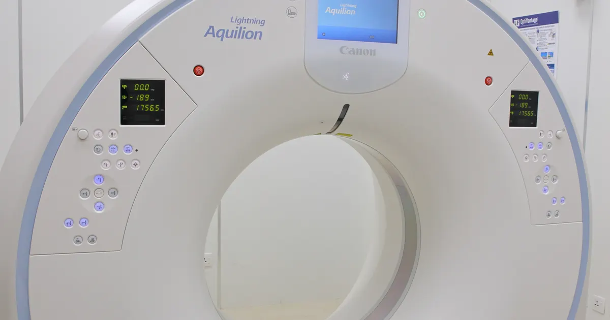
A CBCT scan is not taken for every dental problem — it is only prescribed when the clinical examination and 2D X-rays (periapical, panoramic) do not answer the question and when the 3D information will change the decision. This is not an absolute list, but the logic of indications: bone width and the distance to the nerve/sinus in implant planning (EAO, AAOMR), complex root canal treatment and retreatment (the joint AAE/AAOMR position statement), a wisdom tooth close to the nerve in the lower jaw, impacted teeth in orthodontics (AAOMR 2013), bone pathology of the jaw joint, odontogenic sinusitis, trauma, cysts and tumours. The dose of a CBCT scan is far higher than that of a panoramic X-ray (tens to hundreds of µSv depending on the FOV), which is why every scan is justified and the smallest possible field is selected.
The clinical workflow behind these indications and the price are explained on the 3D diagnostics service page.
International guidelines (the European SEDENTEXCT document, the AAOMR and AAE position statements, the EAO consensus) stress that a CBCT scan should be taken on an appropriate and justified indication. Below we explain the 10 most common situations and why a CBCT scan is needed in each of them.
1. Dental implant planning
A dental implant is placed inside the bone; this is why 3D accuracy regarding bone height, width and density and the neighbouring anatomical structures (nerve, sinus) is essential.
- In the lower jaw the distance to the mandibular canal (nerve) is measured — injury is prevented.
- In the upper jaw the height up to the sinus cavity is measured — and a sinus lift is planned if needed.
- Bone width determines the implant diameter.
- DICOM data is needed in order to make a surgical guide (guided surgery) at the clinic.
For details see Why CBCT matters for dental implants and the dental implant service page at our clinic.
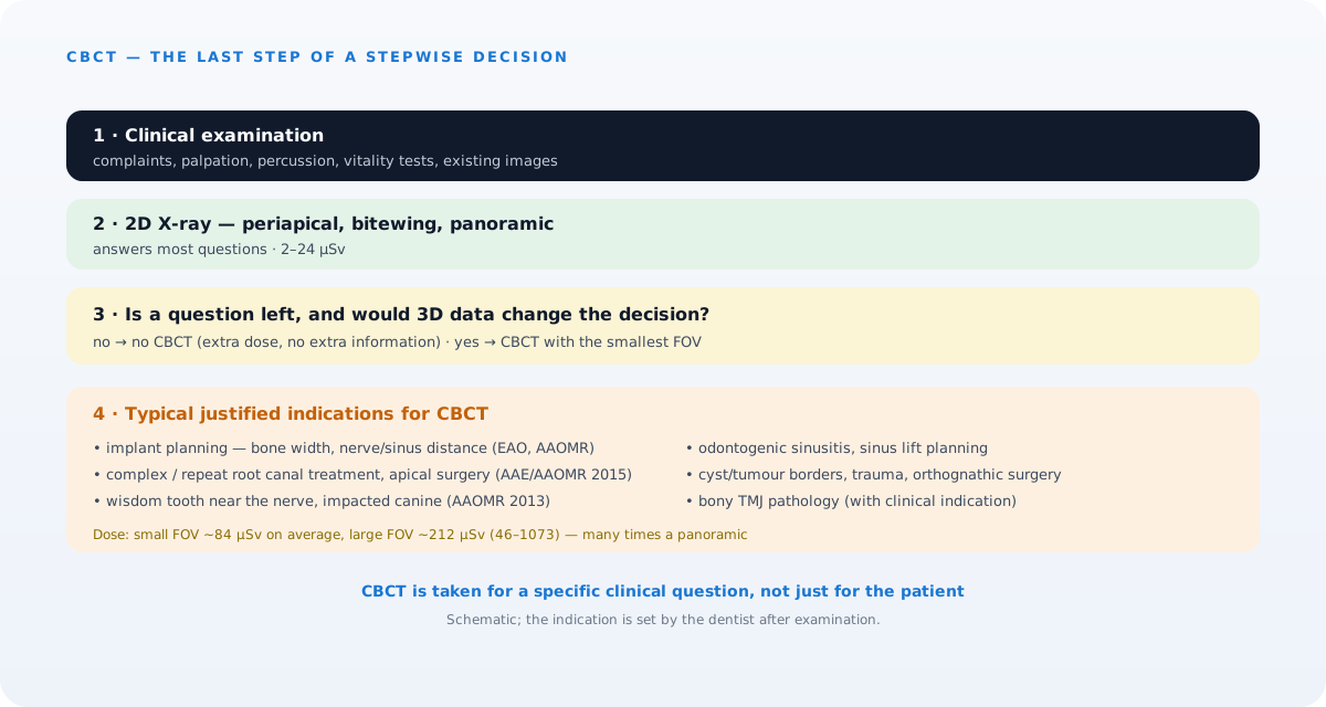 A CBCT scan is the last step of a stepwise decision, not the first one. The diagram is schematic.
2. Difficult root canal treatment (endodontics)
An ordinary X-ray shows only 2 of the 3 dimensions. A CBCT scan reveals the following:
- Extra canals (for example the second mesiobuccal canal in upper molars, which is often not visible in 2D).
- Root curvature and complex internal anatomy.
- Perforation, a fractured instrument or extra material inside the canal.
- Periapical inflammation (cyst / abscess) becomes visible at an earlier stage.
- Resorption (dissolving of the root) is assessed in its true extent.
According to the 2015 joint position statement by the AAE and the AAOMR, a CBCT scan is not routine in endodontics: it is recommended with a small FOV when 2D X-rays are not enough — an unclear diagnosis, complex anatomy, retreatment, planning of apical surgery, root resorption and trauma cases.
3. Wisdom tooth (third molar) removal
A lower wisdom tooth is sometimes very close to the nerve of the lower jaw (inferior alveolar nerve). If the nerve is damaged, numbness of the lip may become permanent. A CBCT scan reduces this risk to a minimum:
- The exact distance between the tooth root and the nerve canal is measured.
- The angle at which the tooth lies and its relationship with the neighbouring tooth root can be seen.
- In the upper jaw, proximity to the sinus is assessed.
- The surgeon plans the surgical approach in advance (tooth sectioning, coronectomy and so on).
4. Orthodontic treatment planning
A CBCT scan is recommended for orthodontic treatment particularly in the following situations:
- Impacted canines — the exact position of the tooth and whether there is any root damage.
- Skeletal asymmetry and planning of orthognathic surgery.
- Assessment of the bone defect in cases of cleft palate / lip.
- Root position and proximity to the gum before treatment with clear aligners and braces.
For routine orthodontics a panoramic X-ray plus a lateral cephalometric X-ray is usually enough; the 2013 AAOMR position statement recommends a CBCT scan in orthodontics only on an individual indication and with the smallest FOV.
5. Jaw joint (TMJ) problems
In cases of painful clicking, limited opening and closing, or a jaw that locks, a CBCT scan:
- Shows the shape and position of the condyle (the head of the joint).
- Detects signs of osteoarthritis (condylar erosion, osteophytes).
- Assesses congenital asymmetry and developmental disorders.
For the soft tissue (the disc) an MRI is needed; where the question concerns bone structure, a CBCT scan is the choice. However, with painless clicking or simple muscle pain, imaging is usually not needed — the diagnosis rests on the clinical examination (TMJ service).
6. Toothache of sinus origin and odontogenic sinusitis
The roots of the back teeth in the upper jaw are often very close to the maxillary sinus (sometimes they enter the sinus). If there is a problem in this area, it is not clear whether the source of the pain is a tooth or the sinus. A CBCT scan:
- Shows thickening of the sinus membrane, fluid, polyps and cysts.
- Detects signs of odontogenic sinusitis spreading from a tooth root.
- Assesses whether an implant or root canal treatment has affected the sinuses.
See our article Can a CBCT scan show sinus problems on this subject.
7. Jaw trauma and fractures
In cases of a road accident, a sports injury or a fall:
- Alveolar fractures and dislocation of teeth.
- Condylar fractures and jaw fragments.
- Complex fractures of the facial bones.
A CBCT scan gives an accurate 3D image and is invaluable for planning surgery. In high-risk cases, medical CT may be preferable.
8. Cysts, tumours and suspicious lesions
A shadow seen on a panoramic X-ray raises questions that a CBCT scan answers:
- What size is the lesion and how far has it spread into the tissues?
- Is its border cortical, or has it passed into the soft tissue?
- Are there hyperdense (hard) parts inside it?
- Where is the access point for surgical removal or a biopsy?
If the spread into the soft tissue is extensive, medical CT/MRI is added. For a list of the conditions that can be seen on a CBCT scan, see the article Which conditions can be seen on a CBCT scan.
9. Jaw surgery (orthognathic and reconstructive)
A CBCT scan is essential in the surgical correction of the lower jaw forwards / backwards, of the upper jaw downwards / upwards, or of asymmetry. Simulation software carries out the planning on the DICOM data and surgical guides are produced with 3D printers.
10. Assessment of aesthetic cases and closed tooth positions
In zirconia bridges, complex aesthetic rehabilitation and restorations close to the gum, the bone structure and the position of the roots are planned with a CBCT scan. In full-arch All-on-4 / All-on-6 plans in particular, the position of the implants is planned on the 3D image (All-on-4 service).
When is a CBCT scan not needed?
- An ordinary dental check-up and an examination for decay — 2D bitewing / periapical X-rays are enough.
- Routine orthodontic monitoring — a panoramic X-ray plus a lateral cephalometric X-ray is enough.
- Examinations that can be postponed during pregnancy — they are postponed.
- A preventive CBCT scan every 6 months — it should not be taken if there is no indication.
Who refers you for a CBCT scan?
A CBCT scan is recommended by your dentist or your maxillofacial surgeon on the basis of the clinical examination and the results of 2D X-rays. In some cases an orthodontist, an endodontist or an ENT doctor may also write a referral. Having the scan on the patient's own initiative is not recommended — without an indication, the dose is not justified.
FAQ — Frequently asked questions
1. Is a CBCT scan needed for every implant?
Not automatically for every implant — the EAO and AAOMR guidelines recommend the clinical examination and the available 2D images first, and then cross-sectional 3D imaging if a question remains. In practice, because the bone width at the implant site and the distance to the nerve/sinus cannot be measured by any other method, a CBCT scan is often justified; if a sufficiently recent image is available, it is not repeated.
2. Is a CBCT scan always needed for root canal treatment?
No, usually not for an ordinary root canal. However, a CBCT scan becomes essential with difficult root anatomy, retreatment, and when the cause of the pain is not clear.
3. If I already have a panoramic X-ray, why is a CBCT scan needed?
A panoramic X-ray is 2D and does not give information about depth in 3D. Bone width, the distance to the nerve and the condition of the sinus cannot be seen fully on a panoramic X-ray.
4. Can children have a CBCT scan?
Yes, but only if there is a serious indication (orthodontic impaction, trauma, cleft palate) and with the smallest FOV. The dose is kept to a minimum.
5. Can a dentist make a mistake based on a CBCT result?
A CBCT scan is a powerful tool, but only an experienced dentist can read it correctly. At our clinic every 3D result is checked by a dentist.
Conclusion and booking an appointment
A CBCT scan is not taken for the patient — it is taken for a specific clinical question. A CBCT scan taken on the right indication increases the accuracy of planning; a scan taken without an indication gives nothing but an extra dose. If you have an implant, a wisdom tooth close to the nerve, complex root canal treatment or a surgical plan, your dentist will tell you after the examination whether a CBCT scan is needed; on the subject of dose, see Is the radiation from a CBCT scan high?.
👉 Book an appointment at our clinic in Baku for a CBCT scan and a treatment plan — 3D imaging and an accurate reading of the results in one place.
Author: Dr. Bakhtiyar Aliyev — dentist, chief doctor of the clinic. This article has been reviewed by a dentist.
Sources
- AAE and AAOMR Joint Position Statement: Use of Cone Beam Computed Tomography in Endodontics 2015 Update — Oral Surgery, Oral Medicine, Oral Pathology and Oral Radiology, 2015
- Position statement of the American Academy of Oral and Maxillofacial Radiology on selection criteria for the use of radiology in dental implantology with emphasis on cone beam computed tomography — American Academy of Oral and Maxillofacial Radiology (OOOO), 2012
- Clinical recommendations regarding use of cone beam computed tomography in orthodontics. Position statement by the American Academy of Oral and Maxillofacial Radiology — Oral Surgery, Oral Medicine, Oral Pathology and Oral Radiology, 2013
- E.A.O. guidelines for the use of diagnostic imaging in implant dentistry 2011. A consensus workshop organized by the European Association for Osseointegration — Clinical Oral Implants Research, 2012
- Effective dose of dental CBCT — a meta analysis of published data and additional data for nine CBCT units — Dentomaxillofacial Radiology, 2015
This article is for general information only and does not replace an individual diagnosis, examination or treatment. Consult a doctor for a decision about your case.