

Jaw Joint (TMJ) Treatment
Jaw joint complaints — pain, clicking, limited opening, morning muscle fatigue — do not always come from the joint itself: the chewing muscles, bruxism, bite and general factors are assessed together. A diagnosis is never based on a single sign such as a click; treatment follows the cause and the clinical findings.
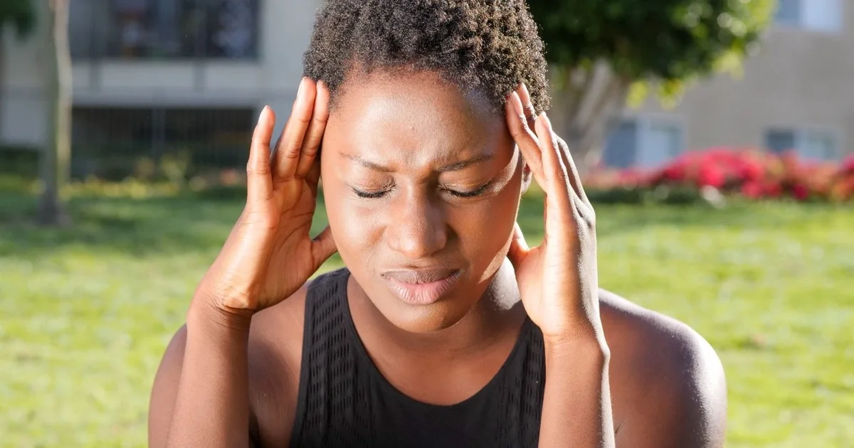
The temporomandibular joint (TMJ) works during chewing, speech and yawning. Complaints in this area are often multifactorial: the joint itself, the chewing muscles, night-time clenching (bruxism), the bite, stress and sleep may all play a part. Our approach is therefore not "one sign — one treatment": complaints and history first, then examination of jaw movements, muscles and joint, bite assessment; imaging and axiography only when clinically indicated; treatment — from self-care to a splint — according to the findings.
The assessment is carried out by orthodontist-gnathologist Dr. Maharram Mammadov. Consultation, functional assessment and appliance prices are listed on the Prices page; the plan follows the examination.
What is a jaw joint (TMJ) problem?
Temporomandibular disorders (TMD) are an umbrella term for pain and functional problems of the jaw joint, the chewing muscles and the structures that connect them: displacement of the joint disc (clicking, sometimes locking), degenerative changes inside the joint, muscle-related pain (myofascial pain) and combinations of these. The international DC/TMD classification bases the diagnosis on clinical examination — the complaint, the location of pain, signs during movement and palpation findings are assessed together. Symptoms, causes and a patient-friendly explanation are in a separate article.
Which symptoms call for an assessment?
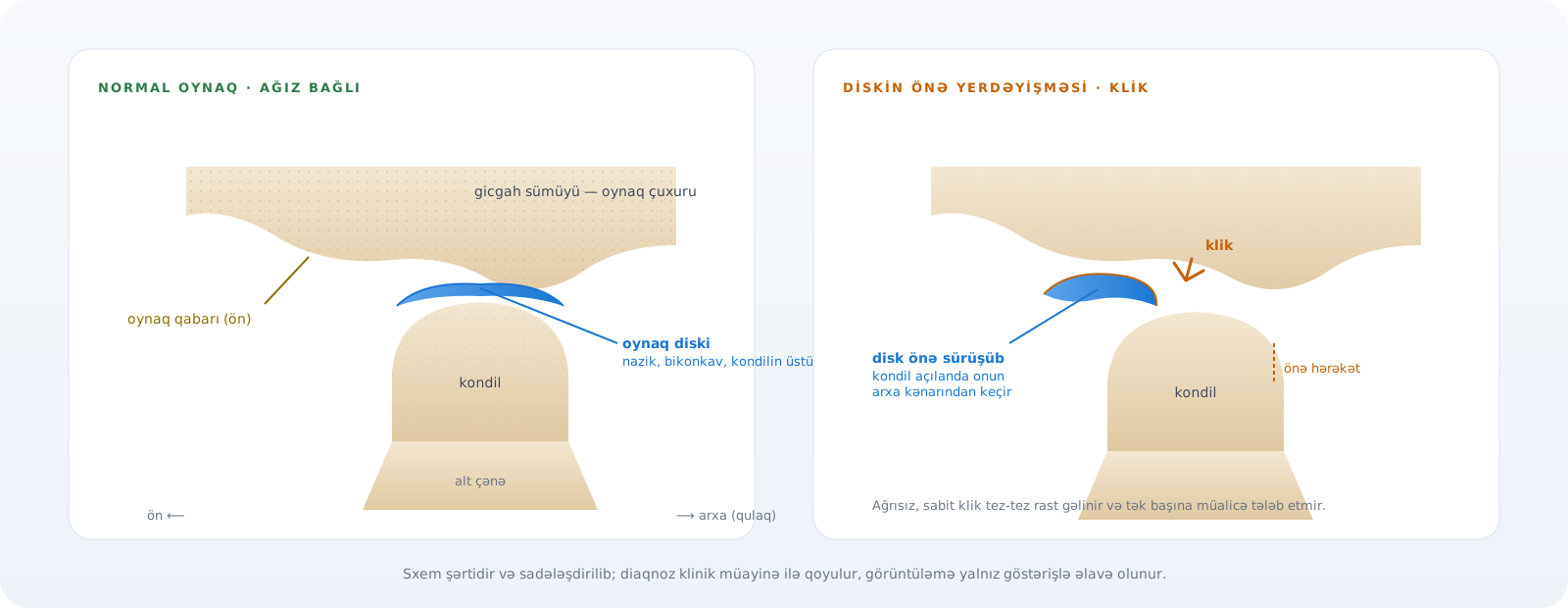
Important: not every click is pathology. A painless, stable click is common in the population and does not by itself need treatment; an assessment is needed when there is pain, impaired function or changing symptoms.
- Pain in front of the ear or in the jaw — worse with chewing, talking or in the morning
- Limited mouth opening, the jaw deviating to one side on opening, or a feeling of “locking”
- Clicking or grating — especially together with pain, catching or impaired function
- Discomfort when chewing, avoiding hard food
- Tired, tense jaw muscles in the morning, headache in the temple area
- Signs of clenching and grinding — tooth wear, scalloped tongue edges, sensitivity
- A feeling of fullness or noise in the ear — once an ENT cause has been ruled out
How is a TMJ problem assessed?
The diagnosis starts with a clinical examination, not with an image. In our clinic the assessment runs in consecutive stages and only the necessary steps are added.
- 1
Complaint and medical history
Location and character of the pain and when it gets worse; history of clicking and locking; sleep, stress, clenching; trauma, previous orthodontic and prosthetic treatment; systemic diseases and medication.
- 2
Clinical examination
Facial symmetry, teeth, wear facets, soft tissues; the range and path of mouth opening (deviation, limitation), lateral and protrusive movements.
- 3
Muscle and joint palpation
Tenderness of the chewing muscles (masseter, temporalis and others) and the joint area; the character and timing of sounds during movement — following DC/TMD principles.
- 4
Occlusal assessment
Tooth contacts, interferences in sliding movements, the wear pattern; models or an intraoral scan when needed. Occlusion is recorded as a finding, not automatically as “the cause”.
- 5
Imaging — only when indicated
CBCT when bony changes are suspected, MRI for questions about the disc and soft tissues; not taken routinely for every patient.
- 6
Axiographic / functional analysis — when needed
Recording the movement paths of the lower jaw: asymmetry, restricted movement and parameters for restorative planning.
- 7
Individual treatment plan
According to the findings: education and self-care, load reduction, a splint, physiotherapy or referral to another specialist when needed; a restorative/occlusal plan only when indicated.
What does axiography provide?
Axiography is a functional analysis method that records the opening, closing, protrusive and lateral movements of the lower jaw. It shows the movement pattern of the joint (including the difference between the two sides), traces that point to the behaviour of the disc during movement, and the individual parameters needed for occlusal planning. It is not a “device that settles the diagnosis 100%” — it complements the clinical examination and is used above all in complex restorations, tooth wear and full-mouth plans.
Imaging: CBCT, MRI — for whom and when?
The great majority of TMJ problems are diagnosed clinically; imaging is ordered when there is a question that would change the outcome. CBCT shows the bony components of the joint — condyle shape, cortical bone, degenerative changes, trauma; MRI is superior for the disc and soft tissues. For a painless click or simple muscle pain imaging is usually unnecessary.
When can splint therapy be used?
A splint (occlusal appliance) is not a universal solution. Its purpose depends on the clinical picture: reducing the load on the muscles and joint, softening the effect of night-time clenching, managing symptoms in some disc problems, or testing the jaw position before a restorative plan. The design also differs by purpose (stabilisation splint, deprogrammer, anterior repositioning appliance). A splint is used under supervision — for a defined period, with periodic adjustments; it is stopped when the complaint resolves or the goal is reached. Systematic reviews and network meta-analyses show that in painful disc displacement conservative approaches (education, exercises, a splint) are often sufficient and invasive interventions are not the first choice.
Bruxism and the jaw joint
Clenching and grinding (bruxism) put parafunctional load on the chewing muscles and the joint; they can appear together with muscle pain, morning fatigue, tooth wear and sometimes joint complaints. International consensus describes bruxism as a behaviour that also occurs in healthy people — it does not cause joint problems in everyone, and not every joint problem comes from bruxism. That is why bruxism is recorded as a finding, the load is managed (a night guard, behavioural advice, sleep and stress factors), and the joint complaint is assessed separately.
Occlusion and gnathology
Occlusion (how the teeth meet) is part of the functional assessment — as a finding, not as the cause. A 2017 systematic review found no consistent cause-and-effect relationship between occlusal features and temporomandibular disorders; the approach “fix the bite and the problem goes away” therefore has no scientific basis, and irreversible treatment of the teeth is not recommended as a therapy for joint pain. The relationship between occlusion and the jaw — centric relation, vertical dimension — is taken into account in tooth wear, multiple restorations and full-mouth rehabilitation plans; in those cases functional analysis and a provisional-restoration stage become part of the plan.
Treatment options — according to the findings
- Education and self-care — soft food, avoiding wide opening and chewing gum, heat/cold, simple opening exercises; in many cases the first and sufficient step
- Load reduction — awareness of daytime clenching, habit change, attention to sleep and stress
- A splint — design and duration according to indication, under supervision
- Physiotherapy or referral to another specialist — for persistent muscle pain, a neck component, or neurological or rheumatological suspicion
- Restorative and occlusal planning — only with a clear indication (tooth wear, missing teeth, full-mouth rehabilitation); not as pain treatment
- Surgery — rarely, for specific joint conditions that do not respond to conservative treatment, together with a maxillofacial surgeon
What we do — and what we do not claim
- TMJ problems can be multifactorial; we do not pin the cause on “one thing”
- Not all clicks need treatment — a painless click is monitored
- Not all pain comes from the joint itself — muscle pain is among the most common causes
- Not every patient needs a splint; a splint does not “put the disc back”
- Not every patient needs CBCT or MRI
- Irreversible changes to the occlusion are not recommended as treatment for joint pain
- No guarantee of outcome; in chronic cases the goal is to restore function and manage pain
Duration and price
The length of treatment depends on the findings: self-care and education can take effect within a few weeks; splint therapy with review visits can continue for months; restorative plans follow their own calendar. The prices below come from the same source as the site’s Prices section; whether an appliance is needed and the overall plan are decided after the examination.
| Initial visit, consultation and personalised treatment plan | 60 AZN |
| Orthodontist examination | 30 AZN |
| SDI Matrix | 600 AZN |
| Kois deprogrammer | 1200 AZN |
| ARA (Anterior Repositioning Appliance) | 1200 AZN |
| Occlusal splint | 1500 AZN |
Prices come from the same source as the Prices page; the final amount is set after your examination. All prices →
Dr. Maharram Mammadov and the gnathological approach
In our clinic the assessment and treatment of TMJ complaints are carried out by orthodontist-gnathologist Dr. Maharram Mammadov, the author of the site’s articles on the jaw joint, splints, bruxism and occlusion. The essence of the gnathological approach is to assess the joint, the muscles and the occlusion as one functional system rather than separately, and to keep intervention as minimally invasive as possible. About our doctors: doctors.
Sources
- Diagnostic Criteria for Temporomandibular Disorders (DC/TMD) for Clinical and Research Applications: recommendations of the International RDC/TMD Consortium Network and Orofacial Pain Special Interest Group — Journal of Oral & Facial Pain and Headache, 2014
- Temporomandibular disorders and dental occlusion. A systematic review of association studies: end of an era? — Journal of Oral Rehabilitation, 2017
- Treatments for painful temporomandibular disc displacement with reduction: a network meta-analysis of randomised clinical trials — International Journal of Oral and Maxillofacial Surgery, 2024
- International consensus on the assessment of bruxism: Report of a work in progress — Journal of Oral Rehabilitation, 2018
- The Role of Imaging in the Diagnosis of Temporomandibular Joint Pathology — Oral and Maxillofacial Surgery Clinics of North America, 2018
If this sounds familiar
If you have one of these complaints, this service may be right for you:
- “My jaw clicks when I open my mouth”
- “My jaw and temples hurt in the morning, I clench”
- “I cannot open my mouth fully”
Frequently asked questions
Is a clicking jaw joint dangerous?
A painless, stable click is common and does not need treatment by itself. Assessment is needed when it comes with pain, locking or limited opening.
Do I need an MRI?
Not everyone. The diagnosis is mainly clinical; MRI answers questions about the disc and soft tissues, CBCT about bony changes.
Who treats jaw joint problems?
At our clinic, orthodontist-gnathologist Dr. Maharram Mammadov; when needed together with a physiotherapist, neurologist or oral surgeon.

Read more on this topic
- Jaw joint problems (TMD): symptoms, causes and when to see a dentist
- Clicking and popping in the jaw joint: what does it mean and when should you worry?
- Splint therapy for the TMJ: what is an occlusal splint and who does it help?
- What happens after splint therapy? Outcome, relapse and next steps
- SDI Jaw Tracker: why is a digital recording of jaw movement needed?
- What is bruxism and how does teeth clenching affect the TMJ?