

When are MRI, CBCT and axiography used for TMJ pain?
With jaw joint (TMJ) pain the first step is not an image but a clinical examination: the dentist checks where the pain is, how far the mouth opens, the joint sounds and the muscles using a standard protocol (DC/TMD). Imaging is taken only when the answer will change the treatment. When a disc or soft tissue problem is suspected, MRI is chosen; when a bone change (arthrosis, erosion, fracture) is suspected, CBCT; and when the way movement is disturbed needs to be documented, axiography. Most patients need none of these.
Key points
- According to the international DC/TMD protocol, muscle and joint pain can be diagnosed reliably by clinical examination alone; imaging is not required for these diagnoses.
- MRI shows the position of the disc and the joint fluid; CBCT does not show the disc, but it shows the condylar surface and bone changes precisely.
- Axiography records movement, not structure; it does not replace MRI, it complements it.
- A panoramic X-ray is only a general overview of the joint; it often misses subtle bone changes.
- Routine CBCT or MRI is not recommended for every TMD patient.
Why the clinical examination first?
Most jaw joint problems (TMD — pain and functional disorders of the joint and the chewing muscles) are of muscular origin, or are pain coming from the joint capsule. For these diagnoses there is an international DC/TMD (Diagnostic Criteria for TMD) protocol published in 2014: the dentist asks where the pain is, presses on the muscles and the joint with a defined force, measures in millimetres how far the mouth opens, and notes clicking and grating.
In the validation project of this protocol (TMD patients and a control group, with expert diagnosis and imaging as the reference standard), the sensitivity of the clinical criteria for muscle pain (myalgia) was 0.90 and the specificity 0.99; for joint pain (arthralgia) they were 0.89 and 0.98. In other words, where the pain is coming from can very probably be identified without an image, through a proper examination. In these patients imaging usually adds no further information.
The situation changes with problems inside the joint. In the same project, the sensitivity of the clinical criteria was only 0.34 for disc displacement with reduction (where the disc returns to place with a click) and 0.55 for degenerative joint disease (arthrosis). This is why the DC/TMD document states: a definitive diagnosis of disc displacement requires MRI, and degenerative change requires CT (including CBCT) — but only where that diagnosis will change the treatment plan or the prognosis.
In plain terms: first the dentist examines you, then chooses imaging according to the question “what will the answer change?”. We have explained separately in signs of a jaw joint problem when symptoms are serious.
Which question — which examination?
The joint can be seen through three different “eyes”: soft tissue, bone and movement. Each method shows one of these well, and the others poorly or not at all.
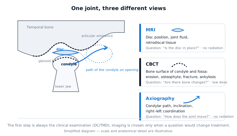 In the same joint, three examinations look at three different structures: the disc (MRI), the bone surface (CBCT), the movement path (axiography).
| Criterion | MRI | CBCT | Axiography | Panoramic X-ray |
|---|---|---|---|---|
| What it shows | Disc position, joint fluid, retrodiscal tissue, bone marrow | Bone surface of the condyle and the fossa, erosion, osteophytes, fracture, ankylosis | Path of condylar movement, inclination, right-left coordination | General contour of the condyle, major deformity |
| What it does not show | Fine detail of the bone surface | Disc and soft tissue | No structure at all — function only | Disc, subtle bone change |
| Main question | “Is the disc in place, is there inflammation?” | “Is there arthrosis, erosion or a fracture in the bone?” | “How does the joint move, is the movement asymmetrical?” | “Is there anything obviously wrong?” (initial overview) |
| Radiation | None | Yes (low dose) | None | Yes (low dose) |
| Typical indication | Locking together with clicking, limited mouth opening, cases where the MRI result will change the treatment | Grating (crepitus), suspected bone change with long-lasting pain, trauma, surgical/orthodontic planning | Major prosthodontic and orthodontic planning, functional evidence when a disc problem is suspected, treatment monitoring | Initial overview as part of a general dental examination |
MRI: the disc and soft tissue question
MRI (magnetic resonance imaging — a radiation-free method that shows soft tissue using a magnetic field) is the only routine examination that shows the disc in the joint directly. According to the 2023 joint position statement of the American Academy of Oral and Maxillofacial Radiology (AAOMR) and the American Academy of Orofacial Pain (AAOP), when disc displacement is suspected, MRI taken with the mouth both closed and open should be used; CBCT, because of its low soft tissue contrast, cannot assess the disc.
Reading MRI is reliable as well. In a study published in 2009 within the RDC/TMD validation project, four board-certified radiologists read the joint images of 724 participants using standard criteria: on MRI, inter-examiner agreement (kappa) was 0.78 for disc displacement with reduction, 0.94 for displacement without reduction and 0.64 for joint fluid (effusion). In other words, when read with standard criteria, two radiologists largely reach the same conclusion on the same MRI.
When is MRI needed? The DC/TMD answer is practical: when the disc diagnosis will change the treatment. For example, long-standing clicking on its own does not require MRI — many people have clicking in the joint and it is left untreated. But if the clicking is combined with locking, if mouth opening has decreased, if the first conservative treatment has not helped, or if surgery/arthrocentesis is being discussed, knowing the position of the disc changes the decision. You can read what clicking means in sound from the jaw joint.
CBCT: the bone change question
CBCT (cone-beam computed tomography — a low-dose method that shows bone in three dimensions) shows the surface of the condyle (the head of the joint) and of the fossa. The AAOMR/AAOP position statement recommends CT, preferably CBCT, for the bone when arthritis and degenerative change are suspected; a panoramic X-ray is considered insufficient for this purpose, because it does not show most osteophytes and erosions.
In figures: in a systematic review and meta-analysis published in 2016 (8 studies, reference standard — direct visual examination of the condyle, mostly on laboratory specimens), the sensitivity of CBCT for detecting bone defects in the joint was 0.67 and the specificity 0.87. The authors call this “relatively high accuracy”, but add that a negative result does not completely rule out a bone change. In that same 2009 RDC/TMD study, inter-examiner agreement in the diagnosis of arthrosis was 0.16 on panoramic X-ray, 0.47 on MRI and 0.71 on CT — for bone, CT-type imaging has a clear advantage.
When is CBCT needed? Grating (crepitus) may indicate bone surfaces rubbing against each other — this is a classic indication for arthrosis. Trauma, suspicion of a bone cause of limited mouth opening (ankylosis), long-lasting pain that does not respond to conservative treatment, and orthodontic or surgical planning may also require CBCT. At the same time the position statement warns: routine CBCT for every TMD patient is not justified; and an additional scan with the mouth open is not routinely recommended, because it changes the diagnosis in only a small number of cases. We have explained what exactly CBCT shows in the joint in TMJ problems on CBCT, and the method itself in what CBCT is.
Axiography: the movement question
Axiography (a radiation-free examination that records the path of condylar movement with a sensor) does not show structure. It documents how the joint works — the length, shape and inclination of the path the condyle travels when the mouth opens and during forward and lateral movements, and the coordination of the two sides. We have set out its principle and how the examination proceeds separately in what axiography is.
Its place in diagnosis is complementary. In a study published in 2022, computerised axiography was compared with MRI in the same patients: the sensitivity for detecting disc displacement was approximately 85% and the specificity 95% (MRI as the reference standard, for detection of disc displacement). The authors state clearly: without a clinical examination, axiography has no diagnostic meaning in itself.
When is axiography needed? Mainly in two situations. First, major prosthetic work, full-mouth rehabilitation or orthodontic planning — here joint movement is needed for the individual setting of the articulator. Second, as functional evidence before or alongside MRI when a disc problem is suspected, as well as for comparing movement before and after occlusal splint treatment. For simple muscle pain, axiography is usually not needed.
Is a panoramic X-ray enough?
A panoramic X-ray (a 2D image showing all the teeth and the jaws in one frame) is often taken anyway as part of a general dental examination, and it shows the general contour of the joint. A major deformity, fracture or asymmetry may be noticeable. But it is not a separate diagnostic tool for the joint: it does not show the disc at all, and it mostly misses subtle changes of the bone surface. A note on a panoramic image saying “the joint is normal” does not rule out arthrosis; if there is a suspicion, CBCT is needed.
A practical sequence: what should a patient expect?
- Discussion and examination. Where the pain is, when it started, clicking, locking, morning stiffness, clenching. Then palpation of the muscles and the joint and measurement of mouth opening according to the DC/TMD protocol.
- Initial diagnosis and conservative steps. If muscle or joint pain is found, treatment usually starts without imaging: behavioural advice, exercises and, where needed, an occlusal splint.
- Imaging — with a specific question. “Is the disc in place?” → MRI. “Is there a change in the bone?” → CBCT. “How is movement disturbed, what are the joint parameters for the prosthetic plan?” → axiography. Sometimes two examinations are needed together — for example, when grating comes together with locking, CBCT and MRI complement each other.
- Reassessment. If the treatment does not help, or the symptoms change, the question is asked again and imaging is chosen at that point.
In our clinic this sequence is carried out by an orthodontist-gnathologist; the course of the functional examination is set out on the TMJ and gnathology page.
Frequently asked questions
My jaw clicks, but it does not hurt. Should I have an image taken? For clicking that is painless, without locking and without limiting mouth opening, neither MRI nor CBCT is usually needed. The dentist may examine you and recommend monitoring. When clicking is combined with locking or with reduced opening, the question changes.
The dentist prescribed CBCT instead of MRI — is that right? It depends on the question. When grating, trauma or a bone change is suspected, CBCT is the right choice. If the position of the disc is the question, CBCT cannot answer it — in that case MRI is needed. Sometimes both are required.
If the disc looks displaced on MRI, is treatment definitely needed? No. Disc displacement is also found in people without complaints. The treatment decision is made according to the complaints and the function, not the image; the MRI result is read together with the clinical picture.
If axiography is cheaper than MRI and radiation-free, why is it not done for everyone? Because it does not show the disc; it only documents that movement has changed. For a diagnosis it has to be read together with the clinical examination. Its main benefit lies in prosthodontic and orthodontic planning, as well as in monitoring treatment.
Which examination is chosen for the jaw joint in children? In a developing jaw, the indications for examinations involving radiation are set more narrowly. The clinical examination comes first; the disc question is resolved with MRI (radiation-free), and the bone question with CBCT only where necessary. The decision is made individually.
Conclusion
With jaw joint pain, the choice of examination begins not with the question “which device is more powerful” but with “which question is waiting for an answer”. The clinical examination establishes most of the diagnoses. MRI looks at the disc, CBCT at the bone and axiography at movement, and none of them replaces another. A consultation with an orthodontist-gnathologist is useful for assessing your individual situation and choosing the examination you need.
Sources
- Diagnostic Criteria for Temporomandibular Disorders (DC/TMD) for Clinical and Research Applications — Journal of Oral & Facial Pain and Headache, 2014 (Schiffman et al.)
- Recommendations for Imaging of the Temporomandibular Joint. Position Statement from the American Academy of Oral and Maxillofacial Radiology and the American Academy of Orofacial Pain — Journal of Oral & Facial Pain and Headache, 2023 (AAOMR / AAOP)
- Research diagnostic criteria for temporomandibular disorders (RDC/TMD): development of image analysis criteria and examiner reliability for image analysis — Oral Surgery, Oral Medicine, Oral Pathology, Oral Radiology and Endodontology, 2009 (Ahmad et al.)
- The detection accuracy of cone beam CT for osseous defects of the temporomandibular joint: a systematic review and meta-analysis — Scientific Reports, 2016 (Ma et al.)
- Diagnostic use of computerized axiography in TMJ disc displacements — Experimental and Therapeutic Medicine, 2022 (Talmaceanu et al.)
This article is for general information only and does not replace an individual diagnosis, examination or treatment. Consult a doctor for a decision about your case.