

What bone changes does CBCT show in the jaw joint (TMJ)?
CBCT shows the bony components of the jaw joint — the shape of the condyle, the integrity of the cortical bone, signs of osteoarthritis (erosion, osteophytes, sclerosis, subchondral cysts), ankylosis, fractures and developmental anomalies — more accurately than MRI or a panoramic X-ray; the disc, synovial fluid and muscles, however, are not visible on CBCT, and MRI is needed for these. An important point: according to the RDC/TMD imaging criteria, a joint diagnosis is made on clinical examination, and imaging is added only when there is a question that would change the outcome — for painless clicking or muscle pain, a CBCT is usually not needed. The position of the condyle alone is not considered an indicator of disc displacement.
This article is devoted to the bone findings that CBCT shows in the jaw joint: the integrity of the cortical bone, the shape of the condyle, degenerative changes. Which method (clinical examination, MRI, CBCT, axiography) is chosen and when in TMJ pain is covered in a separate guide: When are MRI, CBCT and axiography used for TMJ pain?. A short rule: bone — CBCT, disc and soft tissue — MRI.
What is the TMJ and what problems occur?
The jaw joint is a delicate joint between the condyle of the lower jaw and the joint socket of the temporal bone. The articular disc between them (fibrous cartilage) smooths the movement. The most common problems in the TMJ are:
- Osteoarthritis — wear of the bone structure, pain, clicking.
- Disc displacement — the disc moving out of its position.
- Myofascial pain — of muscular origin.
- Ankylosis — restricted movement.
- Traumatic fractures — condylar fractures.
- Congenital anomalies — hypoplasia, hyperplasia.
Some of these problems are seen with CBCT, others with MRI.
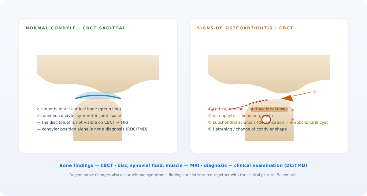 CBCT shows bone findings; the disc and soft tissue are the domain of MRI. The diagram is schematic.
What can be seen in the TMJ on CBCT?
1. Shape of the condyle and bone structure
A normal condyle is rounded and smooth. With CBCT:
- Changes in shape (flattening, deformed shape)
- Wear (erosion)
- Osteophytes (bony outgrowths) — a sign of osteoarthritis
- Subchondral cysts — cavities beneath the condyle
2. Joint socket (glenoid fossa)
The shape and depth of the socket in which the condyle sits.
3. Signs of osteoarthritis
TMJ osteoarthritis is shown on CBCT by the following signs:
- Thinning and disruption of the cortical bone
- Formation of an osteophyte
- Subchondral sclerosis (increased density)
- Change in condyle shape
- In some cases, a subchondral cyst
The combination of these signs establishes the diagnosis.
4. Condyle position
Whether the condyle sits anteriorly, centrally or posteriorly within the socket is visible on CBCT sagittal slices, but according to the RDC/TMD imaging criteria, condyle position is not a reliable indicator of disc displacement — it also varies widely in healthy people; a disc diagnosis is made with MRI.
5. Ankylosis and restricted movement
Cases where the condyle is fused to the socket by bone (bony ankylosis) are detected with CBCT.
6. Traumatic fractures
Condylar fractures (head, neck, subcondylar) are clearly visible on a 3D image. This is needed for the surgical decision.
7. Congenital anomalies
- Condylar hypoplasia (underdevelopment) — in syndromes such as hemifacial microsomia.
- Condylar hyperplasia — one side of the condyle being larger, with facial asymmetry.
8. Symmetry and comparison
Comparing the right and left sides is easy on CBCT. Asymmetry gives a specific diagnostic direction.
What CBCT does not show in the TMJ
- The articular disc — it is cartilage, a soft tissue. It is not visible on CBCT.
- Whether the disc is in its correct position (disc displacement) — MRI is needed.
- Synovial fluid and signs of inflammation — MRI.
- Synovitis and capsulitis — MRI.
- Soft tissue tumours — MRI.
MRI is required for these.
CBCT vs MRI — which and when in the TMJ?
| Clinical indication | CBCT | MRI |
|---|---|---|
| Bone condition of the condyle | ✅ Ideal | Not sufficient |
| Osteoarthritis | ✅ Early signs | Later stage |
| Disc displacement | Not visible | ✅ Ideal |
| Fracture | ✅ 3D accuracy | Soft tissue injury |
| Ankylosis | ✅ Bone assessment | Soft tissue fusion |
| Congenital anomaly | ✅ Bone | Soft tissue |
| Synovitis / capsulitis | Not visible | ✅ Ideal |
| Tumour | Spread within bone | ✅ Spread into soft tissue |
In practice: in a patient with clicking and pain on opening and closing, first a clinical examination + panoramic X-ray, then, if indicated, CBCT (bone condition) and MRI (disc). A combined approach gives the most accurate diagnosis.
Who is a TMJ CBCT recommended for?
- Painful clicking and popping sounds.
- Restricted mouth opening (trismus).
- Locking of the jaw.
- Congenital or developmental asymmetry.
- Suspected condylar fracture after trauma.
- Orthodontic or orthognathic surgical planning.
- Long-standing headache and jaw muscle pain (complex diagnostics).
Not every TMJ patient needs a CBCT — it is taken when there is an indication.
Osteoarthritis — the problem CBCT detects most often
TMJ osteoarthritis is associated with age, sex, parafunctional load and the individual susceptibility of the joint; a consistent causal link with bite (occlusal) features has not been confirmed in systematic reviews (Manfredini et al., 2017). CBCT shows early bone signs (cortical disruption, small osteophytes) better than 2D imaging; however, the finding is interpreted together with the clinical symptoms — degenerative change without symptoms also occurs. Possible approaches:
- Information, load reduction and self-care
- An occlusal splint where indicated (not a universal solution)
- Physiotherapy
- Anti-inflammatory treatment as prescribed by the doctor
- In rare cases, when there is no response to conservative treatment, a surgical consultation
Assessment and treatment approach: treatment of jaw joint problems.
How is a TMJ CBCT scan performed?
The same as a standard CBCT — 3–5 minutes in total, 15–25 seconds of radiation. For the TMJ, usually:
- Mouth closed position (condyle in the socket)
- Mouth open position (the condyle moves forward)
scanning in both positions is sometimes recommended — to partly assess the dynamic movement of the condyle. Total time in the room is 5–7 minutes.
A step-by-step approach to diagnosing a TMJ problem
- Clinical examination: history, pain, clicking, measurement of movement.
- Panoramic X-ray: initial overall assessment.
- CBCT (if indicated): bone structure.
- MRI (if a disc problem is suspected): disc position and inflammation.
- Physiotherapy consultation and/or assessment by the dentist.
- Surgical consultation (in rare cases).
The order of these steps may change depending on the nature of the patient's complaint.
TMJ CBCT in children and young people
In children and young people the TMJ is still developing. A CBCT is taken in cases where a congenital anomaly or JIA (juvenile idiopathic arthritis) is suspected. If a developmental disorder of the condyle is detected early, the timing of orthognathic correction is chosen correctly.
Advice for patients with a TMJ problem
- When the pain is severe, start with drug-free measures (hot and cold compresses, soft food).
- If there is clicking but no pain, treatment is usually not needed — observation is enough.
- If there is pain and restricted movement, see a dentist.
- Do not use a ready-made night guard without a dentist's assessment — an appliance used without a purpose can change or worsen the situation.
FAQ — Most frequently asked questions
1. Which is better for the TMJ, CBCT or MRI?
The two are complementary. CBCT for bone problems, MRI for the disc and soft tissue.
2. Do I have TMJ osteoarthritis — can it be seen with CBCT?
Early signs (cortical disruption, small osteophytes) are visible on CBCT. The diagnosis is made together with the clinical symptoms.
3. I have clicking — do I need a CBCT?
If there is only clicking, with no pain or restriction, a CBCT is most often not needed. Where there is pain and reduced movement, CBCT and/or MRI may be recommended.
4. Does CBCT show the jaw muscles?
No. Muscles are soft tissue and are not clearly visible on CBCT. Pain of muscular origin (myofascial) is diagnosed by clinical examination.
5. Is CBCT used for a TMJ fracture?
Yes. Condylar and subcondylar fractures are accurately assessed with CBCT, and the surgical decision is based on this.
Conclusion and booking an appointment
Diagnosing a TMJ problem has many levels. CBCT detects the bone structure and signs of osteoarthritis early; MRI is used for the disc and soft tissues. A method chosen on the right indication ensures an accurate diagnosis and successful treatment.
👉 Book an appointment at our clinic in Baku for TMJ diagnosis and treatment — CBCT, clinical examination and, where necessary, a recommendation for MRI, all in one place.
Author: Dr. Bakhtiyar Aliyev — dentist, chief doctor of the clinic. This article has been reviewed by a dentist.
Sources
- The Role of Imaging in the Diagnosis of Temporomandibular Joint Pathology — Oral and Maxillofacial Surgery Clinics of North America, 2018
- Research diagnostic criteria for temporomandibular disorders (RDC/TMD): development of image analysis criteria and examiner reliability for image analysis — Oral Surgery, Oral Medicine, Oral Pathology, Oral Radiology and Endodontology, 2009
- Diagnostic Criteria for Temporomandibular Disorders (DC/TMD) for Clinical and Research Applications: recommendations of the International RDC/TMD Consortium Network and Orofacial Pain Special Interest Group — Journal of Oral & Facial Pain and Headache, 2014
- Temporomandibular disorders and dental occlusion. A systematic review of association studies: end of an era? — Journal of Oral Rehabilitation, 2017
This article is for general information only and does not replace an individual diagnosis, examination or treatment. Consult a doctor for a decision about your case.