

What does CBCT show? Which conditions and findings can be detected

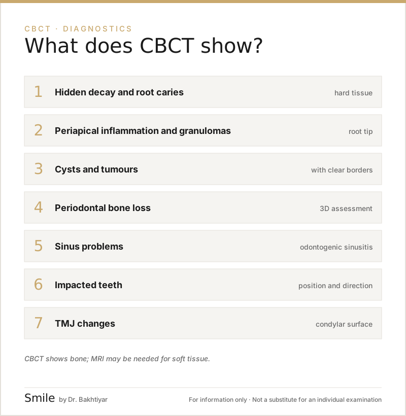
CBCT provides a 3D image of the jaws, teeth, sinuses and the temporomandibular joint. The main conditions it can show are: hidden and hard-to-detect tooth decay, cysts and tumours, periapical abscesses, root canal problems, bone loss (periodontitis), impacted teeth, proximity to the lower jaw nerve, sinusitis, jaw fractures, TMJ osteoarthritis and congenital anomalies. While CBCT is limited when it comes to soft tissue tumours and vascular problems, it clearly shows the vast majority of dental conditions.
What does the research say? According to the joint AAE/AAOMR position statement (2015), CBCT is not a routine scan; it is prescribed to answer a specific clinical question — complex canal anatomy, the extent of periapical inflammation, a suspected root fracture, surgical planning. The AAOMR position from 2012 recommends cross-sectional imaging for implant planning. Systematic reviews show that incidental findings on CBCT (sinus changes, hidden teeth, bone anomalies) are common, that not all of them are ‘disease’, and that they require radiological interpretation. Related: 3D diagnostics (CBCT) service · When is CBCT needed? · What is CBCT?.
This article is about findings — what can be seen on a CBCT image, and what cannot. When a dentist prescribes a CBCT (the indications) is covered in a separate article: In which cases is a CBCT needed?. Below, the findings are grouped into categories — both for the patient and for the dentist who is the first to read the result.
1. Tooth decay and hidden cavities
CBCT is not the first choice for examining tooth decay — a bitewing X-ray and a clinical examination are usually used for this. However, in some cases CBCT makes things easier to see:
- Between the teeth (interproximal) hidden decay that is not visible on 2D images
- Under a crown — recurrent decay
- Root caries (particularly important in older patients)
- Decay of uncertain depth — has it reached the pulp?
Even so, in terms of radiation dose a routine CBCT is not recommended for tooth decay; it is taken when there is a clinical indication.
2. Periapical inflammation (at the root tip)
Conditions located at the root tip appear earlier and more precisely on CBCT:
- Periapical granuloma — a small radiolucent area with a clear border.
- Periapical cyst — a larger area with a thin cortical border.
- Acute and chronic abscess — bone resorption, perforation of the cortical plate.
- Osteitis — widespread inflammation of the bone.
- Osteomyelitis — a rare but serious bone infection.
Especially when the cause of pain is not visible on 2D images, CBCT clearly shows its location and the extent of its spread.
3. Root canal (endodontic) problems
Many conditions related to root canal treatment:
- An unexpected extra canal (for example, the MB2 canal in upper molars is present in around 60% of cases and is not visible on 2D images).
- Root curvature and complex anatomy.
- An isolated perforation — a small hole in the canal wall.
- A fractured canal instrument (a broken endodontic file).
- Vertical root fracture — a crack running along the length of the root; CBCT detects it through indirect signs (localised ‘J-shaped’ bone loss).
- Internal and external resorption — resorption of the root from the inside or the outside.
- Failed root canal treatment (for planning retreatment).
Patients with a root canal problem can visit our Root canal treatment service page.
4. Cysts and tumours (odontogenic and non-odontogenic)
CBCT is invaluable in assessing radiolucent (dark) and partly radiopaque (light) lesions in the jaws:
Benign cysts:
- Periapical (radicular) cyst — at the root tip, inflammatory in origin.
- Dentigerous cyst — around the crown of an impacted tooth.
- Odontogenic keratocyst (OKC) — aggressive, prone to recurrence.
- Residual cyst — remaining at the site of an extracted tooth.
- Nasopalatine duct cyst — in the midline of the upper jaw.
Benign tumours:
- Ameloblastoma — slow-growing, with clear borders.
- Odontoma — made up of dental tissue remnants.
- Cementoblastoma, osteoma — of bone origin.
Suspicious / malignant lesions:
CBCT can sometimes show the bone changes caused by osteosarcoma, metastases and other malignant formations — but MRI / MSCT is needed to assess spread into the soft tissues.
The majority of cysts are benign; so there is no need to panic on seeing the word ‘cyst’ in a report — your dentist will explain it.
5. Periodontitis (bone loss around the gums)
Gum inflammation and bone loss (periodontitis) are very clearly visible on CBCT:
- Vertical bone defects — which tooth, and in which direction.
- Furcation lesions — bone loss where the roots of molars divide.
- Cortical bone loss — in the buccal and lingual directions.
- Crater-shaped defects.
This is important in advanced periodontitis and before a surgical procedure (for example, bone grafting).
6. Sinuses and odontogenic sinusitis
The maxillary sinuses, which lie above the back teeth of the upper jaw, are clearly visible on CBCT:
- Thickening of the Schneiderian membrane — a sign of inflammation.
- Fluid in the sinus — a sign of acute sinusitis.
- Polyps, cysts, retention pseudocysts.
- Odontogenic sinusitis — inflammation spreading from a tooth root into the sinus.
- Root canal filling material passing into the sinus.
- Oro-antral communication — a connection between the sinus and the mouth after a tooth extraction.
For more detail, see our article Can CBCT show sinus problems.
7. Impacted and abnormally positioned teeth
- Impacted wisdom tooth (lower and upper) — its position, angulation and proximity to the nerve.
- Impacted canine — buccal or palatal position.
- Supernumerary (extra) teeth — mesiodens and others.
- Odontoma — dental tissue that has not developed fully.
This information is invaluable for the surgeon and the orthodontist.
8. The lower jaw nerve and anatomical pathways
CBCT shows the structure of the lower jaw in detail:
- The mandibular canal (the pathway of the lower jaw nerve) — critical for implants and wisdom teeth.
- The mental foramen — the opening in the jaw where the nerve emerges.
- Accessory canals — rare, but surgically important.
- Vascular canals — for assessing the risk of bleeding.
9. Temporomandibular joint (TMJ) conditions
- Osteoarthritis — condylar erosion, osteophytes, subchondral cysts.
- Condylar hypoplasia / hyperplasia — developmental anomalies.
- Ankylosis — restricted movement in the joint.
- Traumatic fractures.
- Abnormal condylar shapes.
For soft tissue (the disc), MRI is superior.
10. Jaw fractures and trauma
CBCT provides a precise image in jaw trauma:
- Alveolar fractures — fractures of the bone close to the gums.
- Condylar fractures — in the area of the temporomandibular joint.
- Fractures of the body / ramus / angle.
- Cranio-maxillo-facial complex fractures (limited — MSCT is needed in major trauma).
11. Congenital anomalies
- Bone defects in cases of cleft palate and lip.
- Anodontia — absence of teeth.
- Amelogenesis and dentinogenesis imperfecta — anomalies of the enamel and dentin.
- Syndrome-related jaw asymmetries.
12. Foreign bodies and artefacts
- A fractured canal instrument
- Excess composite in a canal
- Residual material left within the bone
- A metal implant / foreign object
CBCT creates an artefact (a shadow) wherever there is metal, but on modern machines the MAR (metal artefact reduction) filter reduces this.
What CBCT does not show
We should be fair: CBCT does not show some things well:
- Soft tissue tumours (tongue, lip, lymph nodes) — MRI is needed.
- Vascular conditions — contrast-enhanced MSCT is needed.
- Sensory organs (eye, ear, brain) — CT/MRI within the relevant speciality.
- Metabolic bone diseases — DEXA and blood tests.
That is why CBCT is one part of the diagnostic package, not the whole of it.
The radiology report — terms to pay attention to
Phrases frequently found in the final report:
- ‘Clear borders, corticated’ — a sign of a benign lesion.
- ‘Ill-defined border, cortex destroyed’ — further investigation is needed.
- ‘Radiological follow-up recommended’ — a repeat examination in 3–6 months.
- ‘Biopsy recommended’ — referral to a specialist is essential.
FAQ — The most frequently asked questions
1. Can a tumour be seen on CBCT?
Yes, particularly in the jawbone. But MRI is needed for soft tissue tumours. If a suspicious lesion is detected, a biopsy is carried out.
2. Is CBCT needed for tooth decay?
Usually not. A bitewing X-ray and a clinical examination are enough. CBCT is only taken in hidden or complex cases.
3. Can CBCT diagnose sinusitis?
Yes, particularly odontogenic sinusitis (of dental origin). The sinus walls, fluid and membrane thickening are clearly visible.
4. If root canal treatment has failed, does CBCT show it?
Yes. An extra canal, a root fracture, a perforation and the quality of the filling are all detected on CBCT.
5. Is CBCT taken in children with an anomaly?
It is taken when there is a serious indication (an impacted tooth, cleft palate, trauma). The smallest FOV and dose are selected.
Conclusion and booking an appointment
CBCT is one of the most powerful tools in dental diagnostics and detects the vast majority of conditions occurring in the jaws at an early stage. But the result only has value when it is interpreted correctly by an experienced dentist.
👉 Book an appointment at our clinic in Baku for CBCT diagnostics and an explanation of the result — we walk through it together, explaining the result to you and planning your treatment.
Author: Dr. Bakhtiyar Aliyev — dentist, chief doctor of the clinic. This article has been reviewed by a dentist.
Sources
- AAE and AAOMR Joint Position Statement: Use of Cone Beam Computed Tomography in Endodontics — 2015 Update — Oral Surgery, Oral Medicine, Oral Pathology and Oral Radiology, 2015 (AAE/AAOMR)
- Position statement of the American Academy of Oral and Maxillofacial Radiology on selection criteria for the use of radiology in dental implantology with emphasis on cone beam computed tomography — Oral Surgery, Oral Medicine, Oral Pathology and Oral Radiology, 2012 (AAOMR)
- Incidental findings from cone-beam computed tomography in children and adolescents: a systematic review — European Archives of Paediatric Dentistry, 2025
This article is for general information only and does not replace an individual diagnosis, examination or treatment. Consult a doctor for a decision about your case.