

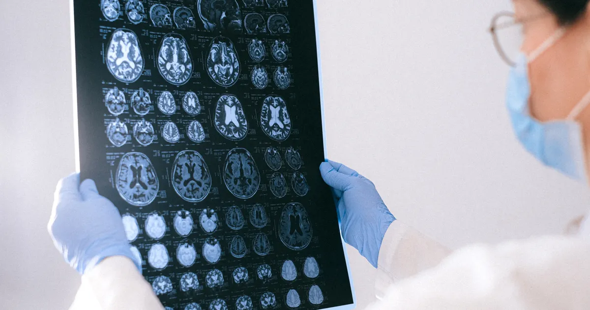
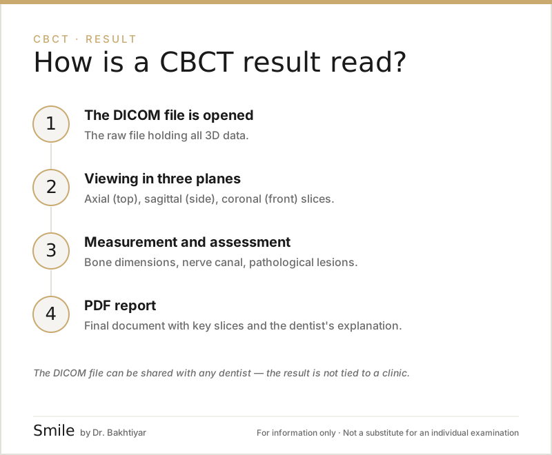
A CBCT result comes in two parts: the raw DICOM file (all the 3D data) and the PDF report (the key slices plus the dentist's explanation). The scan is read in three planes — axial (from above), sagittal (from the side), coronal (from the front) — and in 3D reconstruction. The dentist assesses bone height, the nerve canal, the condition of the sinuses, root anatomy and any pathology.
How the dentist turns these results into a treatment plan is described on our 3D imaging service page.
This article is written for patients: we explain in plain language what each element of the result means, which measurements matter and what you will see in the PDF.
What files make up a CBCT result?
After the scan you are usually given two copies:
- A DICOM file package — the raw 3D data. It may be hundreds of files or a
.ziparchive. - A PDF report — an initial or detailed reading prepared by a dentist or radiologist.
- In some cases: a DVD, USB stick or cloud link.
Any dentist or surgeon can open the DICOM in their own software (for example, planning software). The PDF, meanwhile, provides the clinical interpretation. When you take your results away from the clinic, you should take both with you.
Three main planes — one combined view
Reading a CBCT means using three planes at the same time. Each one answers a different question:
Axial plane (view from above)
This is the slice you get looking down from the top of the head. It shows the cross-section of each tooth.
- The number of canals and the canal anatomy are identified.
- Bone width is measured (for implants).
- The borders of a cyst or tumour are located.
Sagittal plane (view from the side)
This is a slice through the head from right to left.
- The distance between the lower jaw nerve canal and the tooth root is measured.
- The relationship between the sinus floor line and the upper back teeth is visible.
- The angulation of a wisdom tooth (how it is tilted) is assessed.
Coronal plane (view from the front)
This is the plane that slices the head from front to back.
- Sinus asymmetry is detected.
- The height of the lower jawbone and its condition in the bucco-lingual direction are visible.
- The TMJ (temporomandibular joint) can be compared side to side.
3D reconstruction
Using the three planes, the software creates a volumetric model of the whole head. This is used for overall assessment, for producing a surgical guide and for explaining things to the patient.
What does the dentist look at on a CBCT?
During a general reading, the dentist or radiologist assesses the following step by step:
1. The number and condition of the teeth
- Is every tooth present, are any missing, or are there extra (supernumerary) teeth?
- Are there any impacted teeth (teeth that have remained under the gum)?
- Are the tooth roots curved or normal?
2. Bone height and width
These are the key measurements for implants and orthodontic treatment. They are measured in millimetres:
- Vertical height (up to the sinus or the nerve canal)
- Horizontal width (in the bucco-lingual direction)
- Bone density (Hounsfield unit / voxel value)
3. The lower jaw nerve (mandibular canal)
The exact path of this nerve, which runs beneath the lower back teeth, is traced. This is essential for implants and wisdom teeth. A minimum distance of 2 mm from the nerve must be maintained.
4. The sinus cavities
For the maxillary sinus above the upper jaw:
- The thickness of its walls
- Thickening of the membrane (Schneiderian)
- The presence or absence of fluid
- Polyps or cysts
5. Canals and endodontic anatomy
- The number and direction of the canals
- Calcification (canals closing up)
- Areas of resorption
- Perforations, broken instruments
6. Periapical areas (at the root tip)
- Is there a radiolucent (dark) area? → Inflammation, cyst
- What is the border like?
- Has the cortical bone been breached?
7. Anatomical features
- Symmetry of the temporomandibular joint
- Vascular canals, the mental foramen (where the nerve exits the jaw)
- Congenital structures above the teeth
To read more about which conditions can be seen on a CBCT, take a look at our article which conditions can be seen on CBCT.
What will you see in the PDF report?
A typical CBCT PDF report consists of these sections:
- Patient details and scan parameters — name, date, FOV, dose (mSv), voxel size.
- Panoramic reconstruction — an image similar to a 2D view, derived from the 3D data.
- Key slices in three planes — for the area where the problem is.
- Additional images — implant planning, a wisdom tooth section, a sinus section.
- The radiologist's or dentist's interpretation — in written form.
- Recommendations — the next step, if further examination is needed.
A simple report glossary for patients
Here is a plain-language explanation of the terms you will come across most often in your results:
- Radiolucent area — a dark patch; usually means a cavity, inflammation or a cyst.
- Radiopaque area — a light or white patch; hard structure, calcification or metal.
- Cortical bone — the hard outer layer of the bone.
- Trabecular bone — the spongy inner part.
- Periapical — the area at the tip of the root.
- Furcation — the region where the roots divide.
- Schneiderian membrane — the inner lining of the sinus.
- Sclerosis — hardening of the bone, usually a response to long-term inflammation.
- Resorption — dissolving of bone or root.
- Idiopathic — of unknown cause.
Can patients read the results themselves?
Partly. Some parts of the PDF (the written interpretation, for example) are understandable for patients. However, the clinical decision is the dentist's job: the report may show that bone height is 4 mm, but only a dentist can determine that this means it is insufficient for an implant. That is why you should not skip your consultation with the dentist.
What can you do with the DICOM file?
The DICOM file is part of your medical history:
- If you go to another clinic, bring the file with you — a new scan may not be needed.
- It is used to produce a surgical guide (guided implant surgery).
- It can be imported into orthodontic planning software.
- Keep a backup — both in the cloud and on a USB stick.
When do the results need to be updated?
CBCT is not repeated routinely. A new scan is needed if:
- A new treatment plan is required (for example, a new implant in a different site to a healed one).
- There has been a significant clinical change (infection, trauma).
- 1–3 years have passed and the condition of the bone may have changed.
It is usually not repeated within less than a year unless there is a new clinical reason.
FAQ — Most frequently asked questions
1. Can I open the CBCT myself at home?
There are free programs for DICOM files (RadiAnt viewer or OsiriX, for example), but a dentist is needed for a professional reading. The file can be stored at home.
2. Can the PDF report be trusted?
Yes, but only if it has been signed by a radiologist or an experienced dentist. The report should state the author, their speciality and the clinic.
3. If the report mentions a cyst, should I be worried?
No, there is no need to panic straight away. Most cysts are benign; a clinical examination and a biopsy may be needed. Your dentist will tell you the next step.
4. My bone height is 5 mm — can I have an implant?
The decision is not made on the CBCT result alone. Bone width, bone density, the position of the lower jaw nerve and the patient's general condition are all taken into account. In some cases a sinus lift or bone grafting is required.
5. Can I show the results to another dentist?
Of course. The DICOM and the PDF belong to you. You can also present them to another specialist for a second opinion.
Conclusion and booking an appointment
Reading a CBCT result takes care and experience. The raw DICOM file records your individual features, while the PDF is the dentist's interpretation. At our clinic, every 3D result is checked by a dentist and/or a radiologist and explained to you step by step.
👉 Book an appointment at our clinic in Baku for a professional reading of your CBCT results.
Author: Dr. Bakhtiyar Aliyev — dentist, head dentist of the clinic. This article has been reviewed by a dentist.
This article is for general information only and does not replace an individual diagnosis, examination or treatment. Consult a doctor for a decision about your case.