

What is the difference between a panoramic X-ray and CBCT?
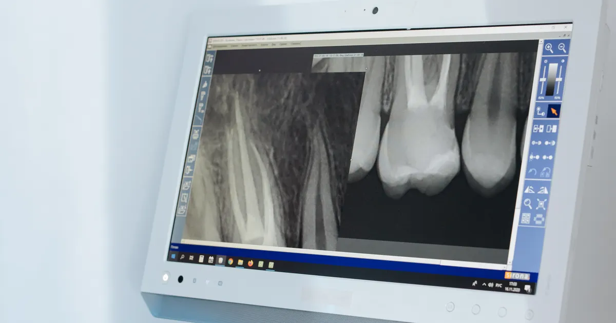
A panoramic X-ray (OPG) is a 2D method that shows the whole jaw in a single image — the dose is low (10–24 µSv), and it is used for a general overview and initial screening. CBCT (Cone Beam Computed Tomography), on the other hand, is a method that creates a 3D volume model of the jaws and teeth — a higher dose (30–1000 µSv), but it allows precise measurement of bone width, the nerve canal, the sinus and implant planning. So an OPG is a map, while CBCT is a 3D model.
Which method is chosen in which situation is explained indication by indication on the 3D imaging service page.
Which X-ray your dentist prescribes for you often raises questions: Why is a panoramic not enough, why is CBCT needed? Or the opposite: Why is CBCT not needed, why is a panoramic enough? In this article I explain the differences between these two methods in terms of use cases, accuracy, radiation dose and price.
Further reading: What is a panoramic X-ray? and What is CBCT (3D imaging)?.
1. Type of image: 2D vs 3D
- Panoramic X-ray: 2D (two-dimensional) — the dental arch is recorded onto a flat plane. Length and height are visible, but there is no depth.
- CBCT: 3D (three-dimensional) — it creates a real volume model of the jaws and teeth. Slices can be taken and viewed in any direction (axial, coronal, sagittal).
This is the most fundamental difference between the two methods. It is like the difference between a paper map and a three-dimensional relief model.
2. How the device works
- OPG: the X-ray tube and the detector make one and a half turns around the head, and successive frames along the face are stitched together.
- CBCT: a cone-shaped X-ray beam is used; during one full or half rotation the device takes projections from hundreds of different angles, and the software converts these into a 3D volume model.
3. Radiation dose
- Panoramic X-ray: approximately 10–24 µSv (microsievert).
- CBCT — depends on the FOV (field of view) and the protocol:
- Small FOV (around one or two teeth, for example 4×4 cm): 30–100 µSv.
- Medium FOV (one jaw): 100–200 µSv.
- Large FOV (both jaws + sinus): 200–1000 µSv.
In other words, CBCT typically delivers 2–40 times more dose than a panoramic (depending on the FOV and the device). Source of the figures: the panoramic and intraoral doses come from the 2008 JADA calculations by Ludlow and co-authors, and the CBCT doses from a 2015 meta-analysis (averages with standard protocols: small FOV 84, medium 177, large 212 µSv; with large differences between devices). Low-dose protocols significantly reduce the dose for the same FOV.
But all these doses are still significantly lower than conventional computed tomography (a head CT is around 1500–2500 µSv). In detail: Is CBCT radiation high?.
4. What does it show?
| Area examined | Panoramic X-ray | CBCT |
|---|---|---|
| All teeth and the dental arch | Yes, the full picture | Yes, in a 3D volume |
| Bone width | No (2D measurement is not possible) | Yes (precise measurement in millimetres) |
| Relationship between the lower jaw nerve canal and the tooth root | General picture, depth not precise | Real 3D distance |
| Sinus floor and pathology inside the sinus | General outline | Detailed 3D |
| Implant planning (length, width, angle) | Initial overview only | Standard tool |
| Interproximal (between the teeth) early tooth decay | Poor | Poor — a bitewing X-ray is needed |
| Accuracy of root canal treatment | General overview | Small FOV CBCT is stronger, but the periapical X-ray is the main tool |
| Suspected cyst or tumour | Initial screening | Detailed border and extent |
| Initial orthodontic overview | Standard tool | Added in complex cases |
| Cephalometric measurement (facial skeletal analysis) | No — a lateral cephalometric X-ray is taken separately | Yes — craniometric measurements can be derived from the 3D data |
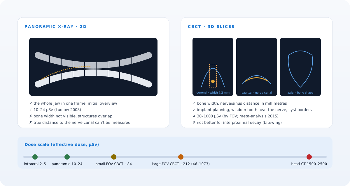 The 2D map versus the 3D model: what each shows and at what dose. The diagram is schematic.
5. Choosing according to the use case
When a panoramic X-ray is more suitable:
- A routine dental examination, a first look.
- Monitoring how permanent teeth are erupting in a child.
- A wisdom tooth decision in a straightforward situation.
- Initial orthodontic screening.
- Initial suspicion of a large cyst or tumour.
When CBCT is more suitable:
- Implant planning (the main indication).
- Complex wisdom tooth surgery (a root close to the nerve canal).
- Sinus lift planning.
- In orthodontics, hidden and impacted teeth, surgical orthodontics.
- Root canal treatment: microcracks, suspicion of a hidden canal, apical surgery.
- TMJ (temporomandibular joint) pathologies.
- Trauma and jaw fracture surgical planning.
- The extent of a cyst, tumour or cancer — the surgical operation plan.
6. Time and process
- Panoramic X-ray: 3–5 minutes including preparation. The exposure takes 15–20 seconds.
- CBCT: 5–10 minutes including preparation. The scan takes 8–40 seconds (depending on the protocol).
Both require you to stay still; neither is painful for the patient.
7. Format of the result
- OPG: usually as a JPEG/PNG or PDF, sometimes DICOM.
- CBCT: a DICOM dataset (a 3D volume made up of hundreds of files). It requires special software (a viewer, for example). The result can be shown to the dentist as a 3D render, as individual slices, and also as a virtual panoramic reconstruction.
For this reason CBCT can in a sense replace a panoramic: a virtual panoramic image can be built from the 3D data. But the reverse is not possible — 3D data cannot be derived from a panoramic.
8. Price
Prices vary from clinic to clinic; at our clinic the current prices are published on the site's Prices page (the panoramic X-ray and 3D imaging are separate lines). The general rule: CBCT is more expensive than a panoramic, because it provides more information and requires longer processing and interpretation — but the choice is made according to the clinical question, not the price.
9. A simple decision scheme
I would like to set out the dentist's way of thinking in a simple form:
- Do you need a first overview? → Panoramic.
- Suspected decay between the teeth? → Bitewing X-ray.
- A deep problem below the root of a single tooth? → Periapical X-ray.
- Implant, complex wisdom tooth, sinus lift, orthodontic surgery, a large cyst, apical surgery? → CBCT.
- A general orthodontic plan? → Panoramic + lateral cephalometric X-ray (sometimes + CBCT).
This scheme reflects the ALARA principle: the right tool for each problem, no more and no less. The ADA's 2024 recommendations follow the same logic — every exposure is justified and optimised.
10. Does CBCT replace the panoramic? Is a panoramic no longer needed?
No. For two reasons:
- Dose: the dose of CBCT is higher than that of a panoramic. For a routine check-up, using a panoramic is the more responsible choice in terms of radiation.
- Price and time: CBCT is more expensive and processing the dataset takes more time. There is no need for that burden for general screening.
That is why modern clinical protocols see these two methods as complementary, not as replacements for one another.
11. A practical scenario: which sequence works when a patient comes to the clinic?
Let us illustrate this with an example scenario. Say a patient complains of frequent pain in the lower right jaw and wants a wisdom tooth taken out:
- Clinical examination — an intraoral look, palpation, percussion.
- Panoramic X-ray — the full picture of the wisdom tooth, the direction of its root and roughly where the lower jaw nerve canal lies become visible.
- If the panoramic shows close contact between the nerve canal and the tooth root:
- A small FOV CBCT is taken — the precise 3D distance of the nerve canal is measured.
- The surgical plan is based on those measurements (for example, coronectomy versus full extraction).
In this scenario it would not be right to reach for CBCT without a panoramic — because there is no point in taking a higher dose without an initial screening test and clinical localisation. At the same time the panoramic alone would not have been enough either — without information about depth, the surgical risk could not have been assessed correctly.
12. The most common misconceptions
- CBCT is better, so it should always be taken. — Wrong. Its higher dose and price cannot be justified for clinically unnecessary use.
- The panoramic X-ray is an outdated method. — Wrong. Digital panoramic devices are one of the main tools of modern dentistry and their dose is very low.
- The CBCT dose is very high, it should be avoided. — When there is an indication for the patient, the dose of CBCT is at an acceptable level given the clinical accuracy it provides (significantly lower than a conventional head CT).
- I had a CBCT once, so I will never need a panoramic. — Wrong. Over time a new clinical indication may arise, and a panoramic may again be suitable for routine monitoring.
Frequently asked questions (FAQ)
Can CBCT replace a panoramic X-ray?
Technically it can do much of the job, but because of its higher dose and price CBCT does not routinely replace the panoramic. It is chosen when there is a clinical indication.
Is CBCT always more accurate than a panoramic X-ray?
Its 3D accuracy is of course higher, but in some situations (general screening, an initial orthodontic overview) a panoramic is the more practical choice and better in terms of dose.
How much more radiation does CBCT deliver?
The dose of a small FOV CBCT is 30–100 µSv, a medium one 100–200 µSv, and a full-mouth CBCT is around 300–1000 µSv. A panoramic, on the other hand, is 10–24 µSv.
Which option is better for an implant?
In implant planning, cross-sectional 3D imaging is often justified for bone width and the distance to the nerve or sinus — the EAO guidelines recommend it after the clinical examination and the available 2D images, when questions remain. The panoramic is for the initial overview.
Which X-ray is better for the orthodontist?
For initial orthodontic planning a panoramic + lateral cephalometric X-ray is required; in complex cases CBCT is added.
About the author
Dr. Bakhtiyar Aliyev — dentist, clinical director of the Smile by Dr. Bakhtiyar clinic. He works in the fields of aesthetic and restorative dentistry, implantology and digital dental radiology. This article has been medically reviewed by Dr. Bakhtiyar.
This article is for general information purposes and does not replace individual medical advice. For a specific diagnosis and treatment, please consult a specialist dentist.
Book an appointment
You can contact our clinic in Baku for panoramic X-ray and CBCT services. Book an appointment online or get in touch with us.
Sources
- Patient risk related to common dental radiographic examinations: the impact of 2007 International Commission on Radiological Protection recommendations regarding dose calculation — Journal of the American Dental Association, 2008
- Effective dose of dental CBCT — a meta analysis of published data and additional data for nine CBCT units — Dentomaxillofacial Radiology, 2015
- Optimizing radiation safety in dentistry: Clinical recommendations and regulatory considerations — Journal of the American Dental Association, 2024
- E.A.O. guidelines for the use of diagnostic imaging in implant dentistry 2011. A consensus workshop organized by the European Association for Osseointegration — Clinical Oral Implants Research, 2012
This article is for general information only and does not replace an individual diagnosis, examination or treatment. Consult a doctor for a decision about your case.