

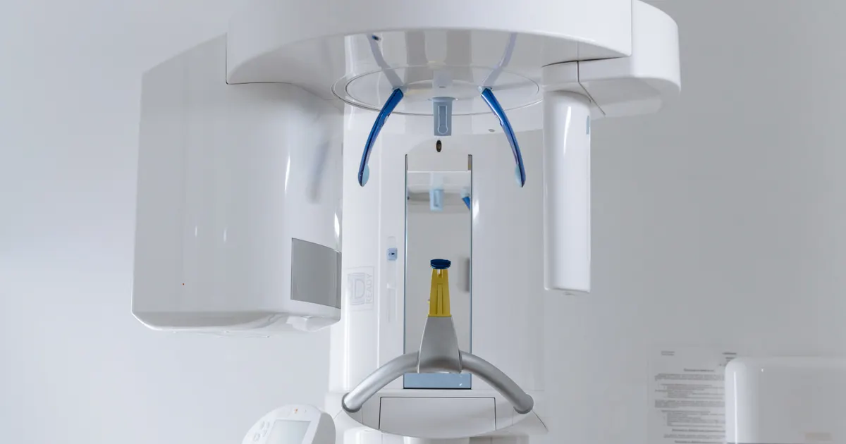
A panoramic X-ray (OPG, orthopantomography) is a 2D dental imaging method that shows the lower and upper jaw, all the teeth, the jaw joints and part of the nasal and sinus cavities in a single image. The unit travels in a half-circle around the head and produces a complete picture of the jaws at a low radiation dose (approximately 10–24 µSv). As a first screening test it is often enough for a general examination, wisdom tooth assessment and orthodontic preparation; for precise 3D measurement (implants), early decay between the teeth and root canal detail, a CBCT, bitewing or periapical X-ray is added.
In the medical literature the panoramic X-ray is known as an orthopantomogram (OPG, OPT). In Azerbaijani it is often simply called a “panorama” or “dental panorama”. In this article I explain in one place how a panoramic X-ray works, what it shows, when it is enough on its own, when it is not, and what its limitations are. Choosing between 2D and 3D is a separate topic: The difference between a panoramic X-ray and CBCT.
How does a panoramic X-ray work?
The patient keeps their head still in a special support (a chin rest and a bite block between the teeth), while the X-ray tube and the detector rotate in a half-circle around the head. During this rotation the unit takes a series of images along the face and combines them digitally into a flat, “unfolded” map. In this way the dentist sees the three-dimensional dental arch in a single flat image.
The stages of the procedure
- Preparation: metal items — earrings, necklaces, hair clips, removable dentures, glasses — are taken off.
- Positioning: the head is placed on the chin rest, the teeth are positioned on the bite block, and the unit checks the eye–ear line.
- Exposure: over 15–20 seconds the unit makes a half-turn around the head; during this time you are asked to stay still, keep the tongue against the palate and hold your breath.
- The image: on digital units the image appears on the screen a few seconds later.
The whole procedure, including preparation, takes no more than 3–5 minutes.
What does a panoramic X-ray show?
- All the teeth — erupted and unerupted, baby and permanent; the length and direction of the roots.
- The jawbone (upper and lower) and its overall condition.
- The jaw joint (TMJ) — on both sides, in general outline.
- Wisdom teeth — their position, angle of eruption, degree of impaction and approximate proximity to the nerve canal in the lower jaw.
- Cysts, large areas of inflammation, large pathologies.
- The lower borders of the upper jaw sinuses.
- The transition from baby teeth to permanent teeth — in children.
- The general condition of old fillings, root canal treatments and implants.
This range makes it a first screening test: with a single exposure the dentist obtains a general picture of the whole mouth and jaw structure.
When can a panoramic X-ray be enough?
“Enough” means that the dentist obtains all the information needed for a decision from the panoramic image alone. Typical situations:
- The general appointment of a new patient. The number of teeth, large pathologies, the condition of previous treatment — if there is no particular suspicion at the clinical examination, a panoramic X-ray is usually enough for a general profile.
- Initial assessment of a wisdom tooth. The angle of eruption, the direction of the root and pressure on the neighbouring tooth are clearly visible; if the tooth is in a straightforward position, a panoramic X-ray is enough for the decision to extract it.
- Initial screening before orthodontic treatment. The number of teeth, impacted teeth, the baby-to-permanent transition; for a full plan a lateral cephalometric X-ray is also needed (Which X-ray is needed for braces?).
- Initial screening when a large cyst or tumour is suspected. Pathologies such as radicular cysts, dentigerous cysts and keratocysts give a clear outline.
- Initial assessment after jaw trauma.
- Monitoring baby and permanent teeth in children. The number of permanent teeth, resorption of the baby tooth roots, impacted teeth.
- The first stage of implant planning — for a general overview only; the final plan is built with a CBCT.
When is a panoramic X-ray not enough?
A panoramic image is two-dimensional — it does not give depth, bone width or real 3D distances. For this reason a separate method is added in the following situations:
| Clinical question | Panoramic X-ray | Additional method |
|---|---|---|
| General overview | ✅ | — |
| Early decay between the teeth | ⚠️ poor (overlap) | Bitewing |
| Root canal/root problem in one tooth | ⚠️ | Periapical, small FOV CBCT for detail |
| Implant plan — bone width, distance to nerve/sinus | ❌ | CBCT |
| Real distance between a wisdom tooth and the nerve canal | ⚠️ shows overlap, not contact | CBCT |
| Microcrack | ❌ | Small FOV CBCT + clinical tests |
| Exact borders of a cyst/tumour | ⚠️ | CBCT, MRI for soft tissue |
| Pathology inside the sinus, TMJ disc | ⚠️ outline only | CBCT / MRI |
| Soft tissue | ❌ | Clinical examination, biopsy, MRI |
In practice the dentist often uses two methods together: panoramic + bitewing (general overview + decay screening), panoramic + periapical (a deep problem in one tooth), panoramic + lateral cephalometric (orthodontic plan), panoramic + CBCT (implants, complex wisdom teeth, large pathologies). This follows the ALARA principle — the right X-ray for each problem, no more and no less. About bitewing and periapical methods: What is a bitewing X-ray?, What is a periapical X-ray?.
When might a CBCT be needed?
The most common reasons for moving on to 3D imaging after a panoramic X-ray:
- Implant planning — the length and width of the implant and the distance to the nerve canal and the sinus can only be measured correctly with a CBCT; a 2D image can give a magnification error of up to 20–25%.
- Overlap between a wisdom tooth root and the nerve canal — structures that overlap in 2D may be several millimetres apart in 3D; a CBCT is needed to assess the surgical risk and to decide on a coronectomy.
- Surgical planning for a cyst or tumour — exact width, depth and proximity to vascular and nerve structures.
- Suspected sinus and jaw joint pathology — CBCT for the bone component, MRI for the disc and soft tissue.
- Complex endodontic cases — a hidden canal, an apical lesion, a suspected microcrack.
The indications for CBCT are covered in a separate article: When is a CBCT needed?. A comparison of the two methods in terms of dose, price and information: The difference between a panoramic X-ray and CBCT.
The limitations of a panoramic X-ray — why does it not show everything?
Patients often ask: “I have had a panoramic X-ray, does it show everything?” The answer is no. There are three technical reasons:
- A 2D image — there is no depth; bone width and the real distance between the nerve canal and a tooth root cannot be assessed.
- The focal plane — the unit focuses on a narrow zone along the dental arch only; structures outside this zone appear blurred.
- Superimposition — where teeth stand close together, shadows fall across one another; this is exactly why early decay between the teeth is hidden.
A practical example: a patient complains of toothache over a long period and was told at another clinic that the panoramic X-ray was “normal”. A bite test causes pain in one specific tooth; a periapical X-ray shows a small area of inflammation at the root tip (on the panoramic image it was covered by the shadow of the tooth above), and a small FOV CBCT reveals a vertical root fracture. A panoramic X-ray could not have shown this on its own — that is not a weakness, but a practical limitation. When your dentist says that an additional X-ray is needed, it is not groundless insistence but clinical responsibility.
Radiation dose, frequency, pregnancy and children
On modern digital panoramic units the effective dose is approximately 10–24 µSv — equivalent to 2–3 days of natural background radiation or one medium-haul flight. There is no “annual limit” for panoramic X-rays; every exposure is justified by a clinical indication (ALARA), and in a healthy adult patient a preventive panoramic X-ray is usually not needed more often than every 3–5 years. The dose table, the question of “how many times can it be taken” and the protection rules are covered in a separate article: Are dental X-rays dangerous?.
- Pregnancy: elective exposures are postponed; in case of urgent need they can be taken with special protection — Can you have an X-ray during pregnancy?.
- Children: with a paediatric protocol (lower dose, shorter exposure), when there is an indication — Are dental X-rays safe for children?.
What should you do before and after the X-ray?
Before: there is no need to fast; remove metal items (earrings, necklaces, hair clips, glasses, removable dentures); tell your dentist if you think you may be pregnant.
During: place your head on the support, position your teeth on the bite block, do not move while the unit rotates, keep your tongue against the palate and hold your breath.
After: nothing special is needed; the result is assessed immediately by the dentist in digital form.
Having a panoramic X-ray in Baku
Criteria to look for when choosing a good clinic: a digital unit (a considerably lower dose than analogue, with the result on screen straight away), regular calibration and a properly supervised radiology service, and image export in DICOM format — important when you want to show the result to another specialist. Alongside panoramic X-rays, our clinic also offers CBCT, lateral cephalometric and intraoral imaging — dental radiology services.
Frequently asked questions (FAQ)
How long does a panoramic X-ray take?
The exposure itself lasts 15–20 seconds; together with preparation and the result it takes no more than 3–5 minutes.
Is a panoramic X-ray painful?
No. The unit does not touch the body, it simply rotates around the head. The only requirement is to stay still and hold your breath during the exposure.
Do you need to come on an empty stomach for a panoramic X-ray?
No, fasting or any special preparation is not required. Only metal items need to be removed before the exposure.
Is the radiation from a panoramic X-ray dangerous?
The dose of one panoramic X-ray is approximately 10–24 µSv — equivalent to 2–3 days of natural background radiation. When taken on a clinical indication, the risk is very low.
Is a panoramic X-ray enough for a routine dental check-up?
For a new patient appointment and a general overview it is often enough. If early decay between the teeth is suspected, a bitewing X-ray is taken in addition.
Does a panoramic X-ray show tooth decay?
Large, deep decay may be visible, but a panoramic X-ray is not reliable for early decay between the teeth — a bitewing is recommended for that.
Is a panoramic X-ray enough for a wisdom tooth extraction?
In a straightforward case, yes. If the tooth root overlaps the nerve canal in the lower jaw, a CBCT is recommended to assess the real distance and the surgical risk.
Is a panoramic X-ray enough for an implant?
No. It is used for a first overview, but a CBCT is needed for bone width and the distance to the nerve and sinus.
Does a panoramic X-ray show a cracked tooth or a cyst?
Large vertical cracks and the initial appearance of large cysts may be visible; for microcracks and the exact borders of a cyst a CBCT is needed (MRI for soft tissue).
About the author
Dr. Bakhtiyar Aliyev — dentist, clinical director of the Smile by Dr. Bakhtiyar clinic. He works in aesthetic and restorative dentistry, implantology and digital dental radiology. This article has been medically reviewed by Dr. Bakhtiyar.
This article is for general information only and does not replace individual medical advice. For diagnosis and treatment, please consult a specialist dentist.
Book an appointment
You are welcome to contact our clinic in Baku for a panoramic X-ray, a CBCT or a general dental examination. Book online or call +994 55 501 01 07.
Sources
- Optimizing radiation safety in dentistry: Clinical recommendations and regulatory considerations — Journal of the American Dental Association, 2024
- Patient risk related to common dental radiographic examinations: the impact of 2007 International Commission on Radiological Protection recommendations regarding dose calculation — Journal of the American Dental Association, 2008
- X-Rays/Radiographs — Oral Health Topics — American Dental Association
This article is for general information only and does not replace an individual diagnosis, examination or treatment. Consult a doctor for a decision about your case.