

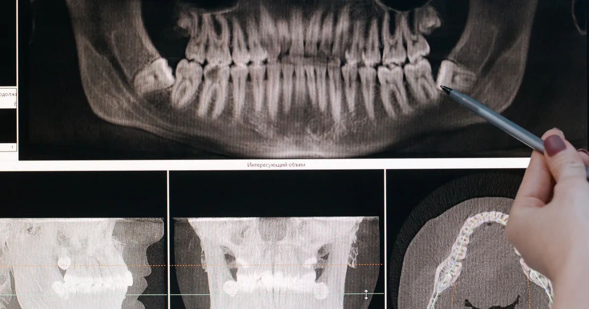
A periapical X-ray is an intraoral (inside the mouth) X-ray image that fully covers one or two adjacent teeth from the crown to the tip of the root (the apex), together with the root canal and the bone structure around the root. A special digital sensor or film is placed inside the mouth, the unit is aimed from the outside, and as a result the dentist sees that area with millimetre accuracy. It is usually used in root canal treatment, deep decay, apical inflammation and suspected root fractures.
What does the research show? A periapical X-ray compresses a 3D structure into 2D: inflammation at the root tip only becomes clearly visible once the cortical bone is affected, so early and small periapical lesions can be missed (International Endodontic Journal, 2009). In the study by Estrela and colleagues (2008), the periapical X-ray failed to show some of the lesions, while CBCT detected significantly more of them; a 2026 meta-analysis also confirms that CBCT shows more lesions in root-treated teeth. Even so, according to the AAE/AAOMR position, the periapical X-ray remains the first choice, with CBCT added only when there is a specific clinical question. Related: Root canal treatment · What is a bitewing X-ray? · What is CBCT?.
Why is a periapical X-ray important?
Panoramic or CBCT X-rays give a broad, overall picture. However, when the dentist wants to see the root tip of one specific tooth, the length of the canal and the area between the root and the surrounding bone at high resolution, the accuracy of a panoramic X-ray is not enough. This is exactly where the periapical (PA X-ray for short) film or sensor is placed parallel to the tooth itself, so angular distortion of the image is kept to a minimum and small changes are clearly visible.
In practice, a periapical X-ray is considered the gold standard for showing the following:
- The length and shape of the root canal (for endodontic treatment);
- Inflammation or the appearance of a cyst at the root tip (the periapical area);
- A root fracture;
- Bone loss beneath the tooth;
- Osseointegration after implant placement (the bonding of the implant to the bone), to monitor it in a small area.

In which cases is a periapical X-ray taken?
In the following situations a periapical X-ray is practically essential:
- When the cause of tooth pain is unclear: to detect decay within the internal structure of the tooth, and especially a hidden lesion where the root joins the crown.
- Before and during root canal (endodontic) treatment: the dentist uses a PA X-ray to measure the working length, to plan for the curvature of the canal and to distinguish between pulpitis and apical periodontitis.
- At follow-up after root canal treatment: to check that the filling material has reached right to the tip of the root and whether there are any voids.
- After trauma: in cases of root fracture caused by a blow, luxation (displacement) of the tooth or damage to the alveolar bone.
- Before a tooth extraction: to assess the shape and curvature of the root and its relationship with neighbouring structures (for example, the lower jaw nerve or the floor of the sinus).
- In implant and periodontal monitoring: to follow the bone level close to the body of the implant and the condition of the periodontal ligaments.
How is a periapical X-ray taken?
The procedure is short and comfortable. It usually takes 1–2 minutes:
- The dentist places a soft holder in your mouth that carries a small sensor or film. The holder keeps the sensor parallel to the axis of the tooth — this is the parallel technique, which reduces distortion.
- The cone (tube) of the X-ray unit is aligned with the external aiming ring of the holder.
- You are asked to hold your breath for a few seconds; the unit delivers the radiation within milliseconds.
- When a digital sensor is used, the image appears on the computer screen immediately. If the image is not clear enough, the dentist can repeat it.
In children, or in patients with a strong gag reflex, a special bite block or an alternative position is chosen; even in this case the radiation dose usually remains very low.
Digital periapical X-ray — why is it better?
In our clinic digital sensors are used instead of film. The reasons for this are:
- Lower dose: a digital sensor requires 50–80% less radiation than film.
- Instant image: there is no need to wait for film processing; the result appears immediately beside the examination chair.
- Enhanced detail: by changing the contrast, brightness and scale, the dentist can see small lesions more clearly.
- Storage in DICOM format: the data goes into the archive in a universal format and can easily be shared with another specialist.
More about digital X-ray technology: What is digital X-ray?.
What does a periapical X-ray show, and what does it not show?
Visible:
- Enamel, dentin, pulp (the internal cavity of the tooth);
- The root canals and their length;
- The periodontal space (the thin gap between the root and the bone);
- A lesion or inflammation at the root tip (radiolucent — a dark area);
- The position of fillings, posts and canal filling materials;
- The level of the alveolar bone (for assessing periodontal disease).
Not visible or limited:
- The sinus, the TMJ, extensive bone pathologies — for these a panoramic X-ray or CBCT (3D imaging) is more suitable;
- A separate view of the buccal (cheek side) and lingual (tongue side) surfaces of the tooth — because it is a 2D image, the structures overlap.
How much radiation is there in a periapical X-ray?
With a single digital periapical X-ray, the effective dose varies roughly between 0.001–0.008 mSv (millisieverts). For comparison, natural background radiation is on average around 0.01 mSv per day. In other words, one PA X-ray is roughly equal to a few hours of natural radiation. That is why, when taken on the correct indication, it is considered safe even in children. More: Is dental X-ray dangerous?.
The difference between a periapical X-ray and a bitewing X-ray
Although both are intraoral X-rays, their purposes differ:
- The periapical X-ray shows the root of the tooth and the bone around the root. For root canal treatment, apical inflammation and trauma.
- The bitewing X-ray shows the crowns of the back teeth of the upper and lower jaw in the same frame. It is used in particular to assess hidden decay at the contact point and early periodontal bone loss.
More: What is a bitewing X-ray?.
Periapical X-rays in pregnancy and in children
During pregnancy, X-rays that are not urgent are postponed. However, if there is severe pain, inflammation or trauma, a periapical X-ray can be taken together with a lead apron (lead cover) and a thyroid collar — because the dose is extremely low, the direct risk to the baby is minimal. In children, a smaller sensor and a shortened exposure setting designed for children are used.
What to do before and after the procedure?
- No special preparation is needed; you do not have to come on an empty stomach.
- Metal items (piercings, glasses, flexible dentures) should be removed from the jaw area — they can create artificial shadows on the X-ray image.
- After the image you can leave the chair and carry on with your normal day; no side effects are expected.
Conclusion: who needs a periapical X-ray, and when?
The periapical X-ray is the dentist's most precise and most frequently used X-ray tool. It is the first choice every time it is necessary to see the canal, root tip and periapical area of one specific tooth clearly: root canal treatment, suspected deep decay, trauma or before a difficult extraction. Its low dose and high accuracy make it reliable for adults and children alike.
Have you been advised to have a periapical X-ray for tooth pain or for a diagnosis? For a modern digital periapical X-ray and CBCT imaging in Baku, you can get in touch with our clinic.
Book an appointment: Online appointment form — you will receive a reply within 24 hours.
Frequently asked questions (FAQ)
1. Is a periapical X-ray painful?
No. The sensor is made of plastic and has soft edges; when it is placed in the mouth you feel only slight pressure. The procedure takes a few seconds and is painless.
2. How many periapical X-rays can be taken in one examination?
When there is a clinical need, 4–8 or more PA X-rays can be taken in a single session (for example, a full-mouth series). The total dose remains well below diagnostic reference levels; every image is justified by an indication.
3. Can decay be seen on a periapical X-ray?
Yes, decay close to the root and proximal (between the teeth) decay in particular is clearly visible. However, for early enamel decay a bitewing X-ray is more sensitive.
4. Is the result given the same day?
The result of a digital periapical X-ray appears on the screen within a few seconds and the dentist goes through it with you there and then. The file can be provided in DICOM or JPEG format.
5. How many periapical X-rays are taken during root canal treatment?
According to the standard protocol, at least three: for the diagnosis before treatment, while the canal length is being measured, and for a check after the filling is completed. Sometimes more X-rays may be required.
Author: Dr. Bakhtiyar Aliyev — dentist, practising in aesthetic and implant dentistry. This article has been reviewed by a dentist (last updated: 2026-07-06).
Sources
- New dimensions in endodontic imaging: part 1. Conventional and alternative radiographic systems — International Endodontic Journal, 2009
- Accuracy of cone beam computed tomography and panoramic and periapical radiography for detection of apical periodontitis — Journal of Endodontics, 2008
- Influence of Imaging Modality on the Detection of Periapical Lesions in Root-Filled Teeth: A Systematic Review and Meta-Analysis — International Endodontic Journal, 2026
- AAE and AAOMR Joint Position Statement: Use of Cone Beam Computed Tomography in Endodontics — 2015 Update — Oral Surgery, Oral Medicine, Oral Pathology and Oral Radiology, 2015 (AAE/AAOMR)
This article is for general information only and does not replace an individual diagnosis, examination or treatment. Consult a doctor for a decision about your case.