

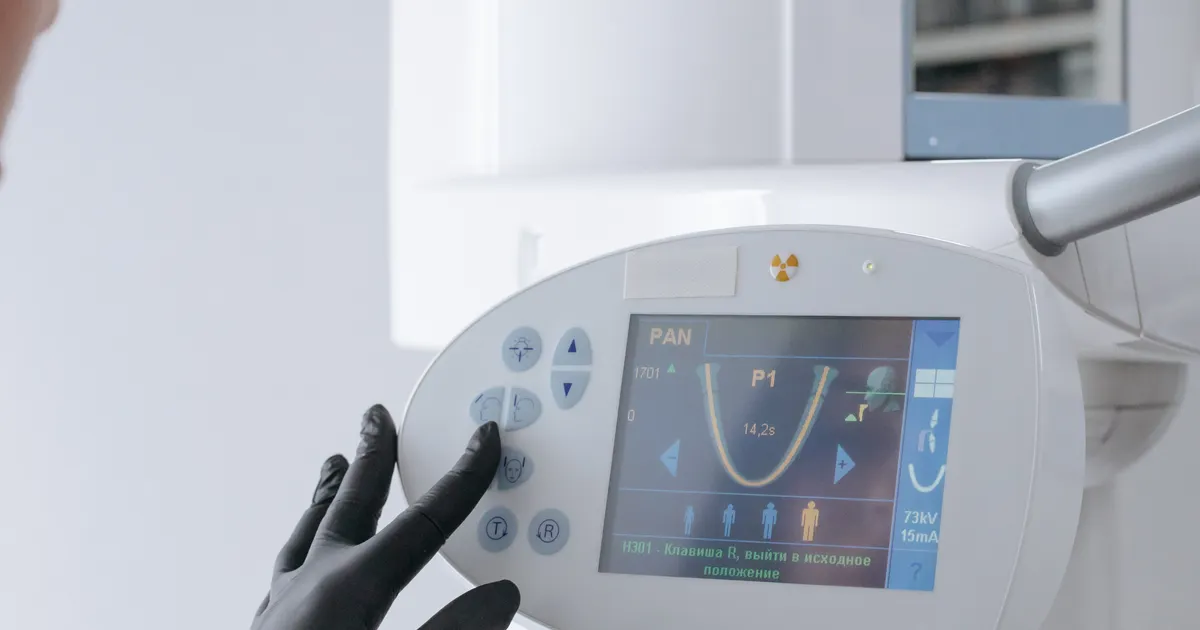
Digital X-ray is a modern imaging method that replaces traditional X-ray film with an electronic sensor. The sensor converts the X-rays into a digital signal immediately, and the image appears on the computer screen within a few seconds. There is no longer any need for film processing, chemical solutions or a darkroom. As a result, the radiation dose drops by 60–80%, diagnosis is faster, the image is stored in DICOM format and can easily be shared via the cloud.
How does digital X-ray work?
The main components of a digital X-ray system:
- X-ray source (tube head): emits the X-ray beam. The power of the X-ray generator is adjusted together with the exposure time.
- Sensor (detector): a CCD/CMOS or phosphor plate (PSP) sensor placed behind the tooth. When the rays reach the sensor, they generate an electronic signal.
- Image processor: converts the signal into a digital image.
- Diagnostic software: displays the image on screen and offers functions such as contrast, brightness, scale, zoom and marking of micro-decay.
Every stage of the process is electronic — there is no film processing at all.

Types of sensor
- CCD (Charge-Coupled Device): the most widely used in intraoral X-rays. The image appears instantly. Because it is wired, it can create positioning limitations in some situations.
- CMOS (Complementary Metal-Oxide-Semiconductor): similar to CCD, but with lower energy consumption. Modern intraoral sensors are mainly CMOS.
- PSP (Photostimulable Phosphor Plate): a plastic plate the size of a film. After exposure it is placed in a special scanner. It has no cable; in some cases it is more comfortable, but it does not give an instant image.
The same principle applies in digital panoramic, cephalometric and CBCT machines — the sensor is simply larger.
Digital vs film X-ray — the main differences
- Dose: a digital sensor requires 60–80% less radiation than film.
- Speed: film required 5–15 minutes of processing; a digital image is ready in about 5 seconds.
- Environmental cost: no chemical solutions, no film packaging, no waste.
- Diagnostic accuracy: contrast adjustment, scale, inverted contrast (invert) and micro-lesion markers give the dentist more information.
- Archiving: it is stored as a file format (usually DICOM, sometimes JPEG) and can still be opened years later.
- Sharing: it can easily be sent to specialists by USB, e-mail or cloud (Google Drive, clinic portal).
- Patient comfort: sensors have softer edges than film and sit comfortably inside the mouth.
See also: Are dental X-rays dangerous?.
The clinical benefits of digital X-ray
- Early diagnosis: by increasing the contrast on the computer, micro-decay, early apical inflammation and micro-fissures can be detected.
- Explaining the image together with the patient: the image can be enlarged and the patient sees the problem with their own eyes — this builds trust and improves treatment compliance.
- Long-term monitoring: previous and current images can be compared side by side; periodontal bone loss or the growth of a cyst is measured objectively.
- AI (artificial intelligence) support: modern software plays a supporting role for the dentist in marking decay, apical pathology and bone level on the X-ray.
- Fast referral: a DICOM file can be sent to an orthodontist, endodontist or surgeon within a few minutes. This speeds up treatment.
How low is the radiation from a digital X-ray?
A comparison framework:
- Digital periapical X-ray: ~0.001–0.008 mSv;
- Digital bitewing X-ray: ~0.005 mSv;
- Digital panoramic X-ray: ~0.02 mSv;
- CBCT (small FOV, low dose): ~0.03–0.1 mSv;
- Conventional film periapical: ~0.005–0.015 mSv (roughly twice as high).
In other words, digital X-ray means both a lower dose and higher diagnostic accuracy.
DICOM and the future of the file
Digital X-ray images are mainly stored in DICOM (Digital Imaging and Communications in Medicine) format. DICOM is not just an image — it carries both the image and metadata (patient, device, date, dose, technical parameters). This allows another dentist, or an implant planning program, to access the file safely and precisely in the future.
Digital X-ray is part of the digital workflow
Digital X-ray does not stand alone; it is the first link in the clinic's digital chain: the DICOM image is combined with the STL model from the intraoral scanner, the implant is planned in software, and the crown is produced with CAD/CAM. For details of the devices and software we use at our clinic: our technologies.
What equipment does a clinic need for digital X-ray?
A fully digital dental clinic usually has the following:
- An intraoral digital sensor (periapical, bitewing and occlusal sizes);
- A digital panoramic machine (sometimes with a cephalometric arm);
- A CBCT system (with a choice of small and medium FOV);
- Image management software (PACS or the clinic PMS);
- A patient portal or cloud sharing service.
Thanks to this integration, the patient can access all of their X-rays, together with their full treatment history, at any time.
Conclusion
Digital X-ray is one of the everyday tools of modern dentistry. Low dose, fast results, high accuracy and easy sharing have closed the film era. In our modern digital X-ray room in Baku, periapical, bitewing, panoramic and CBCT images are taken on a single platform — and the result is given to you both on screen and as a DICOM file.
Book an appointment: Online appointment form
Frequently asked questions (FAQ)
1. Is digital X-ray less harmful?
Yes. It requires 60–80% less radiation than film X-ray. And the diagnostic quality is higher.
2. Will I be given the digital X-ray as a file?
Yes. At the clinic, files in DICOM/JPEG format are given to you by USB, e-mail or cloud sharing.
3. Do I need to come on an empty stomach for a digital X-ray?
No. The procedure is short and simple; no special preparation is needed.
4. Is the appearance of a digital X-ray better than a film X-ray?
Yes — contrast, brightness and scale can be adjusted electronically, which is an advantage in detecting micro-lesions early.
5. What should I ask my dentist about digital X-rays?
Is a digital sensor used?, Is the dose low?, Can I receive the result as DICOM? — these three questions are enough.
Author: Dr. Bakhtiyar Aliyev — dentist. This article has been reviewed by a dentist (last updated: 2026-07-06).
This article is for general information only and does not replace an individual diagnosis, examination or treatment. Consult a doctor for a decision about your case.