

DICOM (Digital Imaging and Communications in Medicine) is the international standard for storing medical images (X-ray, CT, CBCT, MRI, ultrasound) together with both the image itself and its technical and clinical data in a single format. Unlike an ordinary JPEG image, every DICOM file combines the image with the patient name, ID, date, device, dose, slice thickness, exposure parameters, position and dozens of other parameters. This ensures the file opens correctly and can be used clinically in any clinic in the world.
Why is DICOM needed?
For many years medical radiology had a problem: every device manufacturer applied its own format, moving a file from one clinic to another was difficult, and important metadata was lost. The DICOM standard was adopted in 1993 and became the “universal language” of radiology. Today, almost every X-ray and CBCT device in the world produces DICOM.
DICOM is especially important in dentistry, because:
- CBCT results consist of dozens or hundreds of slice files;
- Implant planning programs (Blue Sky Plan, coDiagnostiX, Nemotec, ImplantStudio and others) accept only DICOM;
- Orthodontic and orthognathic surgical plans are built on DICOM;
- The patient’s entire X-ray history is archived in one single format.

What does a DICOM file store?
Every DICOM file consists of two parts:
1. Pixel data (the image)
The greyscale image data produced by the X-ray or CBCT device. In the case of CBCT, each slice is stored as a separate DICOM file, and the software stacks them on top of one another to build a 3D volume. The pixel depth is usually 10–16 bit, which widens the possibilities for adjusting the grey tones (contrast).
2. Metadata (header)
The following are written in the file header:
- Patient data: name, sex, date of birth, patient ID;
- Clinical data: the type of image (CBCT, panoramic, periapical), the indication;
- Device data: manufacturer, model, serial number;
- Technical parameters: kV, mA, exposure time, slice thickness, voxel size, FOV;
- Dose: the DAP or CTDIvol value (if the device records it);
- Date and time: the moment the image was taken;
- Unique file ID: SOP Instance UID (unique worldwide).
Thanks to this metadata, the file opens in any compatible program at the correct scale, contrast and orientation. Most importantly, DICOM preserves the true physical size of the image — how many millimetres each voxel corresponds to. That is why the distance to the nerve canal, bone height and root length can be measured in millimetres within the software.
DICOM is not a single file
With a CBCT scan, DICOM is usually a folder containing hundreds of slices: 200–600 files, 50 MB – 3 GB in total (depending on the FOV and voxel size), files with a .dcm extension or no extension at all, plus a DICOMDIR index. The planning software stacks these slices to build axial (from above), coronal (from the front) and sagittal (from the side) slices, a panoramic reconstruction and a 3D volume model.
The difference between DICOM and JPEG
- JPEG: the image only. It is a compressed (lossy) format. The file size is small. There is no clinical data. The scale is not accurate — it cannot be used for implant planning.
- DICOM: image + metadata + accurate scale + greyscale depth. The file size is large. Implant, orthodontic and surgical programs accept only DICOM.
In other words, a JPEG is a “picture”, while DICOM is a “diagnostic document”.
| Feature | DICOM | JPEG / PNG | PDF report |
|---|---|---|---|
| Three-dimensional slices | ✅ | ❌ | ❌ |
| Measurement in millimetres | ✅ | ❌ | ❌ |
| Implant / orthodontic planning | ✅ | ❌ | ❌ |
| Producing a surgical guide | ✅ | ❌ | ❌ |
| Full analysis for a second opinion | ✅ | partly | ❌ |
| Viewing on an ordinary computer | software needed | easy | easy |
The best practice for a patient is to receive both the DICOM file and the PDF report.
Why is DICOM important for dental planning?
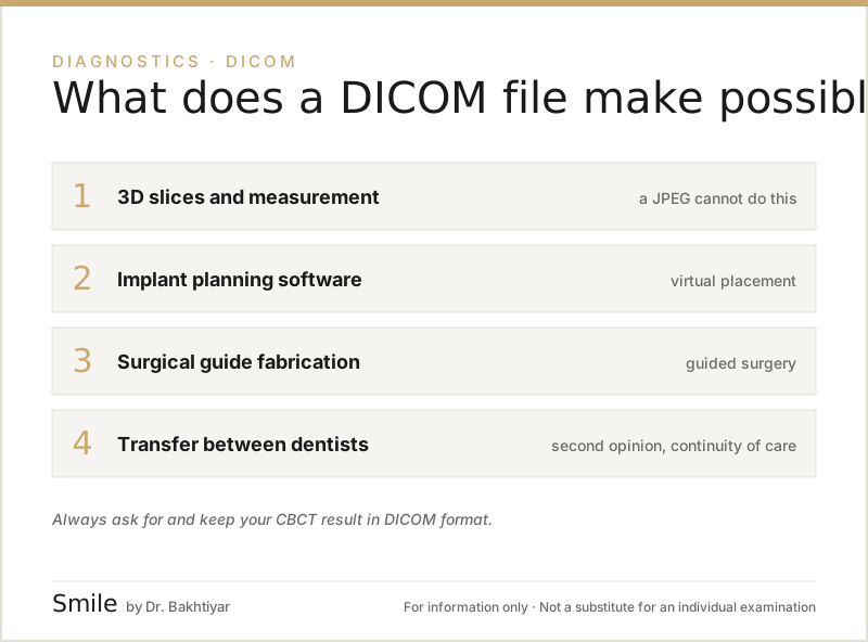
- Three-dimensional slices and measurement. The dentist cuts the volume in any plane and measures in millimetres — the length of the implant, the distance to the nerve and the sinus, the position of the root.
- Implant planning. A virtual implant is selected and positioned in the software, and the distances to the surrounding structures are checked (“computer-guided implantology”). More on this: CBCT and digital planning before an implant.
- Surgical guide. The 3D-printed guide is produced by combining the DICOM data (bone) with the intraoral scan STL (the surface of the teeth).
- Orthodontic and surgical plans. The position of an impacted tooth and the virtual plan for orthognathic surgery are built on DICOM.
- Second opinion and portability. When a patient has the DICOM data, another dentist can analyse the images fully; measurement is not possible on a JPEG or a printout. Because DICOM is a global standard, a clinic abroad can open exactly the same file — which is useful for dental tourism and international consultations. On transferring your results: Taking your X-ray results to another dentist.
What is lost when there is no DICOM?
If the patient is given only a printout or a JPEG: the dentist often has to request a repeat X-ray (extra radiation), the implant plan cannot be built accurately, a surgical guide cannot be produced (freehand surgery is chosen instead), the second opinion is limited, and digital orthodontic planning becomes impossible. For this reason, always ask the clinic for the original DICOM data; it can be stored as a ZIP file or in the cloud, but avoid having it converted into “compressed JPEG” format.
Which programs work with DICOM?
The DICOM viewers and planning programs we use most often at our clinic:
- RadiAnt DICOM Viewer (Windows) — freely available and fast;
- Horos or OsiriX (macOS) — popular DICOM viewers;
- Blue Sky Plan — implant planning (a free version is available);
- coDiagnostiX — implant and surgical planning suite;
- Invivo6, Nemotec — with a dental focus;
- 3Shape Implant Studio, exocad — work with both DICOM and STL together.
If you do not have suitable software on your computer, most of these offer a free demo; and at the clinic the results are always interpreted together with you.
In what format is DICOM given to you?
In dentistry, results are usually handed over in the following ways:
- CD/DVD: the traditional format, supplied together with a ready-to-use viewer program. As some computers no longer have a disc drive, this is gradually falling out of use.
- USB flash drive: the most widely used way of sharing. The files are kept in a single folder together with the
DICOMDIRfile and an accompanying viewer. - Cloud sharing: via a clinic portal, WeTransfer, a Google Drive link or dedicated DICOM storage services (for example dentalcloud.io). This makes sharing with a remote specialist easier.
The type of medium is of secondary importance — what matters is that the original DICOM data is preserved. More on this: Taking your X-ray results to another dentist: format, storage and validity.
Is a DICOM file secure?
A DICOM file carries personal medical data — the patient name, date of birth and ID are shown in it. For this reason:
- Share the file only with a specialist you trust;
- When a patient asks for a file relating to themselves, the clinic cannot refuse to provide it;
- Avoid sharing it through open links on the internet;
- The clinic should keep its DICOM archive on an encrypted server with restricted access.
- For scientific purposes or an international consultation, the file can be anonymised at the patient’s request.
How does DICOM differ from STL?
An STL file is a 3D model file (a three-dimensional surface mesh format). It is obtained from an intraoral scanner, or from the segmentation of a CBCT scan. DICOM, on the other hand, is the tomographic volume and the X-ray image. A detailed comparison: The difference between STL and DICOM.
Conclusion
DICOM is the format standard that forms the foundation of modern medical imaging. In dental practice, CBCT and digital X-ray results are stored and shared precisely as DICOM. The metadata inside it is what makes many planning programs able to work. When sharing files with other dentists, always ask for DICOM — JPEG images and PDF extracts are not diagnostically sufficient.
To receive your CBCT and digital X-ray results as DICOM in Baku, you are welcome to contact our clinic.
Book your appointment: Online appointment form
Frequently asked questions (FAQ)
1. Can I open a DICOM file on my phone?
Yes. There are apps such as “Radiant Lite” for Android and “iDoc” for iOS. However, a desktop viewer is recommended for clinical accuracy.
2. Is a DICOM file large?
A CBCT result can vary between 100 MB and 3 GB. Panoramic and periapical DICOM files are usually 5–30 MB in size.
3. Is the clinic obliged to give me the DICOM data?
Yes, a patient has the right to request a copy of their own medical records. Providing only a JPEG/PDF extract is not enough — DICOM is needed for implant planning.
4. Is DICOM viewer software free?
Many DICOM viewers are free (RadiAnt Lite, Horos, DentiMax Free viewer). Planning programs (implant, orthodontic) usually require a licence.
5. How do I avoid confusing a DICOM file with a JPEG image?
A JPEG is simply a picture of the X-ray image. DICOM is the original volume and metadata. For clinical work, always ask for DICOM.
6. They export the CBCT as JPEG — is that not enough?
It is not enough for implant or orthodontic planning; it is only useful for a quick look. DICOM is needed for measurements and slices.
7. Can another dentist also use my DICOM file?
Yes. DICOM is designed for second opinions and for transferring to another clinic — a dentist anywhere can analyse the same file in full.
Author: Dr. Bakhtiyar Aliyev — dentist. This article has been reviewed by a dentist (last updated: 2026-07-06).
Sources
- About DICOM: Overview — DICOM Standards Committee / NEMA
This article is for general information only and does not replace an individual diagnosis, examination or treatment. Consult a doctor for a decision about your case.