

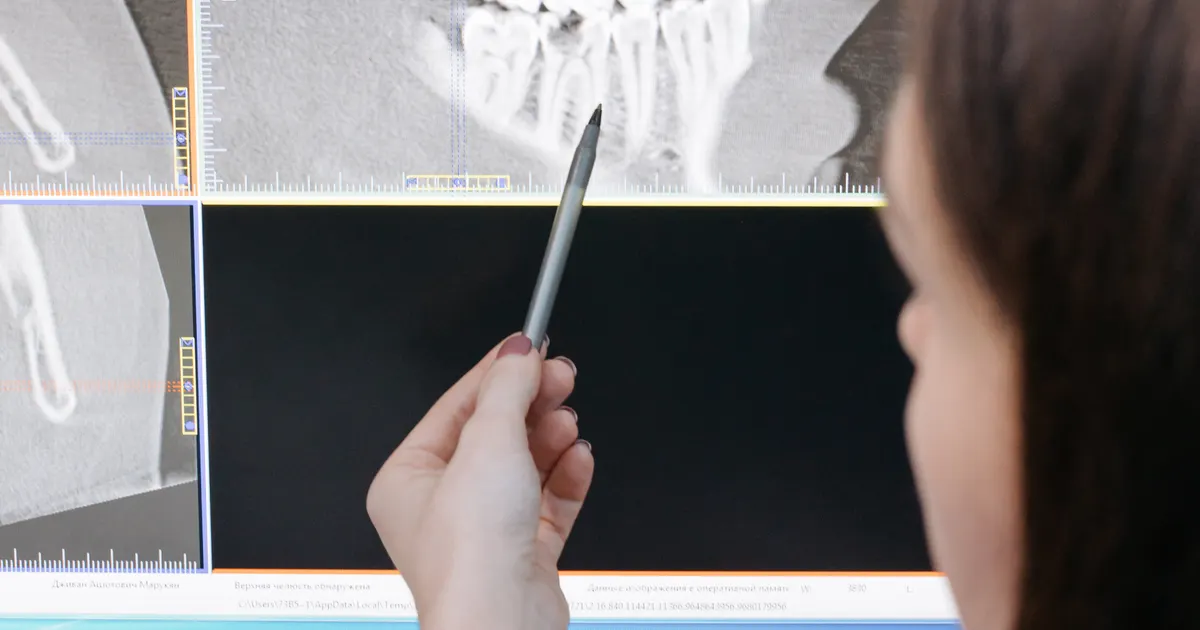
A bitewing X-ray is a type of intraoral X-ray that shows, in a single frame, the crowns of the back teeth (premolars and molars) in the upper and lower jaws, together with the alveolar bone margin of both jaws. The name ‘bitewing' comes from the wing holding the sensor, which the patient bites between their teeth. This image is the most sensitive everyday tool for detecting interdental (proximal) decay, hidden lesions under old fillings and early bone loss. This article is about the technique and the imaging method — how it is taken, which area it shows, when it is chosen; how decay appears on the image and how it is detected is covered in a separate article: Can hidden tooth decay be seen on an X-ray?.
What does the research show? A meta-analysis of diagnostic studies on proximal (interdental) decay reports that bitewing X-rays show high specificity for lesions that have reached the dentin, but limited sensitivity at the early enamel stage — in other words, an X-ray does not show every early lesion, and a negative result does not mean ‘no decay'. Recurrent decay at the margin of a filling is also assessed with the X-ray alongside the clinical examination (Journal of Dental Research, 2016). According to the ADA guideline of 2018, early, non-cavitated lesions visible on an X-ray can be managed with fluoride and sealants rather than an immediate filling — which is why bitewings are used both to detect decay and to choose the level of treatment. Related: Tooth decay treatment · Can hidden tooth decay be seen on an X-ray? · What is a periapical X-ray?.
Why is a bitewing X-ray especially important?
The surfaces where the back teeth rest against one another — known as the contact point — form an area the dentist's eye cannot see directly. Decay that develops in this region (proximal decay) spreads beneath the enamel for a long time without causing any symptoms. To the eye the tooth looks healthy, while inside the cavity may already have reached the dentin. This is exactly where the bitewing X-ray comes in: because the sensor stays perpendicular to the X-ray beam at the point where two teeth meet, the enamel–dentin border is seen without distortion and micro-lesions are picked up.
A digital bitewing X-ray also:
- Makes it possible to measure the height of the alveolar bone — used for the early diagnosis of periodontitis and gingivitis;
- Shows recurrent decay beneath old metal, composite or ceramic fillings;
- Can detect early internal enamel decay that has only just appeared and is not yet visible on examination.

When is a bitewing X-ray taken?
It is usually recommended in the following situations:
- At a preventive check-up: in adults it is advised once every 12–24 months, depending on the risk of decay.
- In children, during the baby and permanent tooth stages: to detect early proximal decay at times when the teeth are very tightly spaced.
- At a new patient examination: to assess previous treatments and the condition beneath existing fillings.
- When planning periodontal treatment: to assess early bone loss and tartar deposits.
- Before orthodontic (braces) treatment: to check whether there is hidden decay between the teeth that are to be moved.
In patients at high risk of decay (a sugary diet, dry mouth, previous fillings) bitewing X-rays may be taken more often, while in low-risk patients they may be taken once every 2–3 years.
How is a bitewing X-ray taken?
The bitewing procedure is extremely simple and takes less than a minute:
- A small digital sensor is placed in the mouth together with a special bite block.
- The sensor is held between the back teeth in such a way that the crowns of both the upper and the lower teeth fall within the same frame.
- The cone of the unit is aligned from the cheek with the aiming ring of the sensor.
- A few seconds of exposure — and the image appears on the computer screen.
Usually two or four bitewing frames are taken, one for the right and one for the left premolar–molar region. In teenagers and children a smaller sensor is used.
What exactly does a bitewing X-ray show?
The structures clearly visible on the image are:
- The crowns of the upper back teeth;
- The crowns of the lower back teeth;
- Enamel, dentin and the pulp chamber;
- Contact decay (proximal decay);
- The integrity of fillings and any gaps beneath them;
- The alveolar bone level (for periodontal disease);
- Early occlusal problems, seen through a discrepancy with the axis of the affected tooth.
A bitewing X-ray does not show the root tip — which is why, when apical inflammation is suspected, the dentist requests a separate periapical X-ray.
The main difference between bitewing and periapical X-rays
These two X-rays are often confused. A simple formula:
- Bitewing = crown + bone level. For hidden decay and early periodontitis.
- Periapical = crown + root + root tip. For root canals, apical inflammation and trauma.
Both may be needed at a single examination. For a detailed comparison: What is a periapical X-ray?.
What does a bitewing X-ray not show?
A bitewing X-ray is not designed to image the following:
- The length of the tooth roots and apical pathology;
- The condition of the nerve in the lower jaw (the ‘mandibular canal');
- The sinuses and the TMJ;
- Bone thickness for an implant — for this a CBCT (3D imaging) is needed;
- Severe decay between the front teeth — because a bitewing is mainly for the premolar–molar region.
Digital bitewing X-rays — the advantages
In our clinic digital sensors are used instead of film:
- A dose 60–80% lower — described in detail in our article What is a digital X-ray?;
- An instant result — discussed while the patient is still in the chair;
- High contrast — electronic filters for detecting micro-decay;
- A DICOM/JPEG file — given to the patient on a USB stick or shared via the cloud.
Is a bitewing X-ray dangerous?
The effective dose is around 0.005 mSv — less than the natural background radiation of a single day. For children a separate lead apron and thyroid collar are used. For detailed safety information: Are dental X-rays dangerous?.
How often is a bitewing X-ray needed?
The general recommendations are as follows (American Dental Association and European protocols):
- In adults with a low risk of decay: once every 24–36 months;
- In adults with a high risk of decay: once every 12–18 months;
- In children preparing for orthodontic treatment: once every 6–18 months (according to clinical indication);
- General rule: an X-ray should not be taken as standard prevention without a clinical indication from the dentist. The dose must be kept to a minimum.
Conclusion: why is the bitewing a ‘small but powerful' X-ray?
The bitewing X-ray is a highly specific tool that helps the dentist see where the eye cannot reach. By finding problems such as hidden decay, recurrence beneath a filling and early bone loss at an early stage, it prevents long and expensive treatments. Together with digital technology, the dose is kept to a minimum and the result is instant.
You are welcome to contact our clinic in Baku for digital bitewing and other dental X-rays.
Book an appointment: Online appointment form — we will get in touch with you within 24 hours.
Frequently asked questions (FAQ)
1. Can a bitewing X-ray be done at home?
No. The X-ray unit and the appropriate sensor are only available in a dental clinic. Using them at home is both impossible and dangerous.
2. What should be done if the sensor triggers a gag reflex?
When the sensor is placed too far back, some patients may develop a reflex. In this case a smaller sensor or an alternative parallel technique is chosen. The problem usually passes within a few short seconds.
3. Does a bitewing X-ray show the depth of interdental decay?
Yes. The depth of decay in the enamel, at the enamel–dentin border and in the dentin can be assessed from the radiolucent shadow.
4. From what age can children have a bitewing X-ray?
It can usually be taken from the age of 4–5 with a small child-sized sensor. The indication is given on the basis of the dentist's assessment of decay risk.
5. How many teeth are visible in one bitewing frame?
Usually 3 teeth from the upper jaw and 3 from the lower — the crowns of 6 teeth in total are fully visible. If needed, a series of 4 frames is taken so that all the back teeth on both sides of the mouth can be assessed.
Author: Dr. Bakhtiyar Aliyev — dentist, practising in aesthetic and implant dentistry. This article has been reviewed by a dentist (last updated: 2026-07-06).
Sources
- Systematic review and meta-analysis of diagnostic studies of proximal surface caries — Clinical Oral Investigations, 2021
- Detecting Secondary Caries Lesions: A Systematic Review and Meta-analysis — Journal of Dental Research, 2016
- Evidence-based clinical practice guideline on nonrestorative treatments for carious lesions: A report from the American Dental Association — Journal of the American Dental Association, 2018 (ADA)
This article is for general information only and does not replace an individual diagnosis, examination or treatment. Consult a doctor for a decision about your case.