

Does an X-ray show hidden tooth decay?
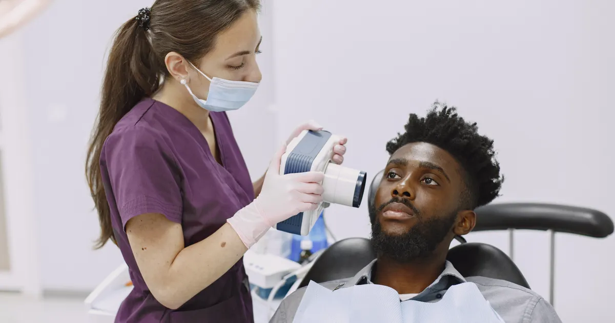
Yes — an X-ray, and the bitewing X-ray in particular, is the most reliable diagnostic method for detecting hidden areas of decay that are invisible to the eye and cause no pain at all. Interproximal decay between the teeth can stay completely hidden during a visual examination, yet on an X-ray image decay shows up clearly as a dark patch. This article is about detecting decay radiographically; the bitewing is explained here as a diagnostic tool, while the imaging technique itself is covered in a separate article: What is a bitewing X-ray?.
Depending on the stage of the lesion that is found, we choose remineralisation, a sealant or a filling — this is part of our tooth decay treatment service.
What is hidden decay and why is it so dangerous?
Tooth decay is a destructive process caused by bacteria that stick to the tooth surface and produce acid, which passes through the enamel and then into the dentin. However, not every cavity troubles you. Hidden decay — decay that the patient does not feel and the dentist cannot see with the naked eye — is the most deceptive kind.
This type of decay most often develops in the following places:
- Between the teeth (the interproximal zone): between the front teeth, as well as between the back teeth. A toothbrush does not reach this area, so decay develops here easily.
- Underneath a filling: the margins of an old amalgam or composite filling lose their integrity over time, and bacteria seep in from underneath.
- At the neck of the tooth: the part where the tooth meets the gum. Decay here usually forms along the gum margin, on the outer surface of the tooth.
- In the deep grooves of the chewing surface: the enamel may look healthy, yet demineralisation may already have started beneath it.
Because it causes no symptoms in the early stage, people often do not see a dentist until severe pain begins. By then, decay may already have reached the pulp, meaning the situation now requires root canal treatment.

Why is the bitewing the gold standard in diagnosing decay?
A panoramic X-ray shows the overall picture — the jawbone, the sinuses, the roots, the periapical areas. However, a panoramic image does not show the area between the teeth accurately enough, because the teeth are projected on top of one another.
The bitewing X-ray, on the other hand, is based on a completely different principle:
- The film (or digital sensor) is placed inside the mouth, next to the teeth.
- The patient bites down on a small plastic holder with their teeth — hence the name bitewing (the wing held between the teeth).
- The X-ray beam passes only through the crown portion of the teeth, that is, through the zone where decay most often begins.
- As a result, the back teeth of both the upper and lower jaw appear in the same image, and the contact point between the teeth is fully visible.
The advantage of this technique is that the beam passing through both sides of the interproximal area reveals the change in density (demineralisation) in that region. Because decay is less dense than enamel, it appears as a darker patch on the X-ray. Your dentist looks at these dark zones to localise the area of decay precisely.
Studies show that a bitewing X-ray can detect interproximal decay 3–5 times earlier than a visual examination.
How does decay look on an X-ray image?
There is a simple rule for reading an X-ray: healthy tissue appears light, that is, white; a cavity, decay or inflammation appears dark, that is, black.
Healthy tooth enamel appears very bright and white on an X-ray. As decay penetrates the enamel surface, that brightness decreases and the area becomes darker. By looking at the size, depth and shape of this dark patch, the dentist determines the stage of the decay:
- Stage D1 (enamel only): a very faint darkening, sometimes visible only on careful inspection.
- Stage D2 (extending into the dentin): the darkening is now clearly visible and reaches up to one third of the depth in the image.
- Stage D3 (extending beyond half of the dentin): decay of this kind is already close to the pulp, and treatment is unavoidable.
Decay underneath a filling is identified by paying attention to the dark halo around the margins of the filling. This can only be seen on a bitewing image.
Our digital intraoral X-ray service makes it possible to carry out this analysis with very high accuracy.
In which cases is CBCT more appropriate?
Although the bitewing is ideal for diagnosing decay, in some more complex situations CBCT (cone-beam computed tomography) is preferable:
- Detailed study of root canal anatomy
- Assessment of periodontal bone loss between multi-rooted teeth
- Planning of implant placement
- Three-dimensional imaging of cysts, tumours and anomalies
However, CBCT is not needed for routine diagnosis of hidden decay. A bitewing X-ray is a faster and cheaper solution with a lower radiation dose. Our CBCT service is used only when there is a clinical indication.
How harmful is an X-ray for decay? There is no need to worry
The question patients ask most often is: is having an X-ray going to harm me?
Modern digital dental X-ray units deliver 70–80% less radiation than traditional film-based equipment. The dose of a single bitewing X-ray is approximately 0.005 mSv — equivalent to just a few hours of the natural background radiation you receive every day (cosmic rays, the soil, building materials).
For comparison:
| Source of radiation | Dose (mSv) |
|---|---|
| One bitewing X-ray | ~0.005 |
| Flight from Baku to Istanbul | ~0.01 |
| Chest X-ray | ~0.1 |
| Daily background (average) | ~0.008 |
A lead cover is always used for pregnant women; other patients are also given a protective apron. Taking these measures into account, the clinical benefit of an X-ray for decay outweighs the potential risk many thousands of times over.
How often should you have an X-ray for decay?
The frequency of X-rays is determined by your individual risk profile:
- Low risk (good oral hygiene, use of fluoride, no history of decay): one bitewing every 18–24 months.
- Moderate risk (patients who sometimes eat a lot of sweets, or have reduced saliva flow): once every 12 months.
- High risk (active decay, dry mouth, immune problems, diabetes): once every 6 months.
Children develop decay faster than adults, so the paediatric protocol calls for more frequent imaging.
For a professional dental examination in Baku, your dentist will determine your risk profile at the first visit and draw up a personalised X-ray plan.
Frequently asked questions
Q: Does an X-ray show hidden decay? A: Yes. Bitewing X-ray images in particular can detect interproximal decay between the teeth at an early stage. Decay that is invisible to the eye and causes no symptoms appears on an X-ray as a dark patch.
Q: How often should I have an X-ray for decay? A: Patients in the moderate risk group are advised to have a bitewing X-ray once a year, and those in the high risk group once every six months. Your individual risk assessment should be carried out by your dentist.
Q: What is a bitewing X-ray? A: A bitewing X-ray is a special intraoral X-ray technique in which the patient bites down on a film holder between the teeth. This method is the most reliable way of detecting interproximal decay between the teeth in particular.
Q: Is it possible to identify decay without an X-ray? A: Large, open areas of decay can be seen during a visual examination, but hidden decay between the teeth, at the neck of the tooth and underneath a filling can very often only be detected with an X-ray. An examination carried out without X-rays is incomplete.
Q: Is an X-ray for decay harmful? A: Modern digital dental X-ray units deliver a very low radiation dose — a single bitewing X-ray is equivalent to a very small fraction of the natural background radiation you receive during the day. When a lead cover is used, the risk is effectively close to zero.
Author: Dr. Bakhtiyar Aliyev, Dental Radiology Specialist | Smile by Dr. Bakhtiyar, Baku. This article has been reviewed by a dentist.
For a professional dental X-ray in Baku, get in touch with us — book online or give us a call.
This article is for general information only and does not replace an individual diagnosis, examination or treatment. Consult a doctor for a decision about your case.