

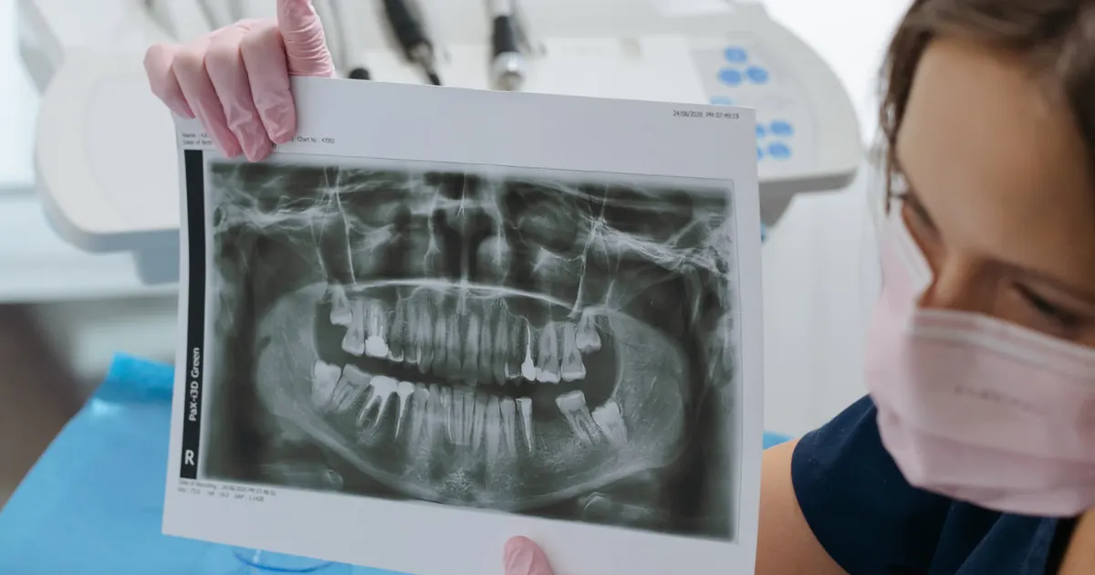
A cracked tooth often does not show up on a standard X-ray. A conventional periapical X-ray can only reveal a fine line when the beam passes exactly parallel to the crack; otherwise the crack stays completely hidden. For a more accurate diagnosis, CBCT (cone-beam computed tomography) combined with a clinical examination is recommended.
Why does a tooth crack not show up on a standard X-ray?
A conventional periapical (small film) X-ray produces a two-dimensional shadow image. As the X-ray beam passes through the tooth, dense structures — the dentin beneath the enamel, the pulp, the root — appear white, while spaces appear dark. A crack, however, does not appear white, because:
- The fine line effect: The width of a crack is measured in micrometres. X-rays pass easily through such a minute gap and leave no trace.
- The direction problem: When the X-ray beam does not fall exactly parallel to the crack, the overlapping tissues mask it.
- Cracks not filled with fluid: Thin cracks containing only air absorb almost none of the X-ray beam.
This situation is known as hidden tooth crack syndrome. The patient is in pain, yet no sign of pathology can be seen on a standard X-ray. That is why the diagnosis of a tooth crack is often delayed.

Which type of X-ray helps most?
Periapical (Small Film) X-ray
This is the most commonly used type. It shows the tip of the tooth and the surrounding bone. Its reliability for detecting cracks is low — especially with vertical cracks. Even so, it is sometimes possible to see a periapical dark area, which can be an early sign of a root crack.
Click here for detailed information about periapical X-rays.
Panoramic X-ray
This covers the whole jaw in a single image. It provides a general overview, but its sensitivity for detecting a crack in an individual tooth is even lower than that of a periapical X-ray, because the image is less detailed.
Bitewing X-ray
This is used for decay between the back teeth. It is of limited use in diagnosing cracks.
CBCT — Cone-Beam Computed Tomography
This is by far the most powerful imaging tool available for diagnosing tooth cracks. CBCT produces three-dimensional slice images (axial, coronal, sagittal). It offers these advantages:
- It can detect very fine cracks running through the tooth.
- It determines the direction of the crack (horizontal, oblique, vertical).
- It shows whether the crack extends as far as the root — information that is decisive for the treatment plan.
What is a CBCT scan? — in this article you will find a detailed explanation of the technology.
Important note: Even CBCT does not reveal 100% of cracks. Very fine vertical cracks may not be visible on CBCT either. For this reason, the X-ray result must always be assessed together with the dentist's clinical examination.
Why is a vertical crack particularly difficult to see on an X-ray?
A vertical crack is the most difficult situation in X-ray diagnosis. A vertical crack runs along the length of the tooth — from the crown towards the roots. Because X-rays usually reach the tooth horizontally, they almost never fall exactly parallel to a vertical crack. As a result, the crack does not appear on the image.
When dentists suspect a vertical crack, they turn to the following additional methods:
- Transillumination: A high-intensity light is held against the side of the tooth. The crack interrupts the light and creates a shadow.
- Dye test (methylene blue): A special dye is applied that shows whether a crack is present.
- Bite pressure test: The patient is asked to bite in different directions; a cracked tooth produces a specific pain.
- Examination under magnification: The specialist checks the tooth using binocular magnification.
Combining these methods significantly improves the diagnosis of a crack.
Cracked tooth syndrome: what are the symptoms?
These are the most common complaints in patients with a hidden tooth crack:
- Sudden sharp pain when chewing — especially with hard foods.
- Pain that disappears suddenly when you stop chewing — this is the characteristic sign of cracked tooth syndrome.
- Excessive sensitivity to cold and heat — the pain lasts longer than a few seconds.
- Night-time pain — the tooth sometimes starts to ache spontaneously.
- Difficulty identifying which tooth hurts — the pain sometimes spreads to several teeth.
If you have these symptoms, waiting is not advisable. As the crack progresses, root canal treatment or even extraction of the tooth may become necessary. Visit our website for information about our dental X-ray service.
Treatment options once a crack has been diagnosed
Treatment differs depending on the depth and direction of the crack:
| Type of crack | Treatment |
|---|---|
| Superficial enamel crack | Polishing, sometimes sealing |
| Cusp crack | Placement of a crown |
| Dentin crack (not reaching the pulp) | Crown, sometimes a seal |
| Crack reaching the pulp | Root canal treatment + crown |
| Vertical root crack | Usually extraction of the tooth |
This is why early diagnosis is of decisive importance. In Baku, a CBCT scan with modern CBCT equipment makes it possible to obtain a complete picture of the crack.
Frequently asked questions
Q: Can a cracked tooth be seen on a standard periapical X-ray? A: Usually not. A standard periapical X-ray can only show cracks that run parallel to the tooth itself. Vertically oriented cracks, or fine cracks not filled with fluid, do not appear on an X-ray. This is why a CBCT scan is recommended for diagnosing cracked tooth syndrome.
Q: Does CBCT always detect a cracked tooth? A: Compared with a standard X-ray, CBCT offers significant advantages in detecting cracks, but it cannot always show very fine or completely vertical cracks. The dentist assesses the CBCT together with the clinical examination.
Q: What does a hidden tooth crack feel like? A: Typical signs are: sharp pain when chewing, sensitivity to hot and cold, and pain when the tooth is pressed in a particular direction. If you have these symptoms, be sure to see a dentist.
Q: Which examination is more accurate for diagnosing a crack? A: At present, the most accurate imaging method is CBCT. However, the result must always be assessed together with the clinical examination — transillumination, the dye test and physical checking of the tooth.
Q: Where can I have a CBCT scan for a cracked tooth in Baku? A: At the Smile by Dr. Bakhtiyar clinic, both X-rays and CBCT scans are carried out with modern CBCT equipment. To book online, you can visit the services section of our website.
Author: Dr. Bakhtiyar Aliyev, Dental Radiology Specialist | Smile by Dr. Bakhtiyar, Baku. This article has been reviewed by a dentist.
For professional dental X-rays in Baku, get in touch with us — book online or give us a call.
This article is for general information only and does not replace an individual diagnosis, examination or treatment. Consult a doctor for a decision about your case.