

Which X-ray shows the position of a wisdom tooth best?
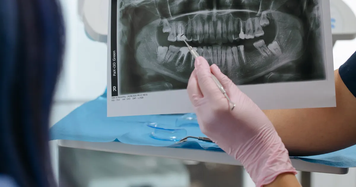
In most cases, a panoramic X-ray is the first and sufficient step for assessing a wisdom tooth: it shows the position of the tooth, its angulation, its relationship with the neighbouring tooth and its overall proximity to the nerve canal. CBCT (cone-beam computed tomography) is added only when risk signs are present — the root overlapping the nerve canal, darkening or narrowing of the canal, deep impaction — in order to refine the surgical plan. CBCT is not needed for every wisdom tooth.
What does the research say? Rood and Shehab (1990) described signs on panoramic X-rays such as the root overlapping the nerve canal and darkening or narrowing of the canal — when these signs are absent, the risk of nerve injury is low and CBCT is usually not required. Comparative studies of panoramic imaging and CBCT report that CBCT shows the root–canal relationship more precisely, but that this changes the surgical plan only in cases with risk signs. NICE guidance recommends removing a wisdom tooth only when there is pathology — imaging is there to support the decision, not to be routine. Related: Oral surgery service · Wisdom tooth extraction · What is a panoramic X-ray?.
Why does a wisdom tooth require special diagnostics?
Wisdom teeth — known by the name that came into dentistry from medicine, the third molars — are the last to erupt and have the greatest potential to cause problems. Very often there is not enough room for them in the jaw. This leads to the tooth remaining partly or completely inside the bone — that is, to wisdom tooth impaction.
An impacted wisdom tooth can cause pain, pressure on the neighbouring tooth, inflammation and even the development of a cyst. To see all these risks in advance, the appropriate X-ray allows the dentist to determine precisely:
- Its depth within the jawbone
- Its angle and direction of tilt
- Its distance from the roots of neighbouring teeth
- Its proximity to the inferior alveolar nerve (nervus alveolaris inferior)
A poorly chosen X-ray method, on the other hand, leaves this information incomplete and creates serious gaps in planning the procedure.

Panoramic X-ray: the first step in wisdom tooth diagnostics
A panoramic X-ray captures all the teeth in both jaws, the jaw joints, the sinuses and the jawbones in a single image. This is by far the most widely used form of wisdom tooth X-ray.
Advantages:
- An overall view of the entire mouth
- A low radiation dose
- A fast imaging process (30–60 seconds)
- It shows both the lower and the upper wisdom teeth at the same time
- Assessment together with the neighbouring teeth, the sinus floor and the jawbone
Limitations:
A panoramic X-ray is a two-dimensional image. This means that it does not show the depth dimension of the tooth — that is, its position in the front-to-back plane. In particular, in the following situations:
- Assessing the full degree of risk for lower wisdom teeth that lie very close to the nerve
- Determining the three-dimensional angulation of the roots
- Calculating the exact distance between the tooth and the bone
a panoramic X-ray on its own may not be enough.
CBCT: the solution for complex wisdom tooth situations
CBCT (cone-beam computed tomography) is the most advanced form of dental radiology. It images the oral and facial region in three planes — horizontal, vertical and front-to-back — at the same time. This is the most important point in the comparison of panoramic imaging and CBCT for wisdom teeth.
When is CBCT needed?
-
Proximity to the nerve: If the root of a lower wisdom tooth appears very close to the inferior alveolar nerve, CBCT shows exactly where the nerve runs. This is critically important for preventing nerve injury during surgery.
-
Deep impaction: If the tooth is positioned deep within the bone, a panoramic image cannot give full information about the condition of the root. CBCT, however, shows every millimetre of the root clearly.
-
Marked curvature and unusual shape: Multi-rooted, hook-shaped root forms, or ones that are not visible on a single X-ray, can be fully identified with CBCT.
-
A problem remaining after a previous procedure: If an attempt was made earlier but the extraction was not completed, CBCT shows the location of the remaining root fragments.
-
Suspected cyst or pathology: When a dentigerous cyst is suspected around a wisdom tooth, CBCT shows its volume, shape and effect on the neighbouring structures precisely.
Let us compare panoramic X-ray and CBCT
| Feature | Panoramic X-ray | CBCT |
|---|---|---|
| Image dimension | 2D (two-dimensional) | 3D (three-dimensional) |
| Overall position of the wisdom tooth | Good | Very good |
| Distance to the nerve | Moderate accuracy | Very high accuracy |
| Root shape | Limited | Complete |
| Radiation dose | Low | Moderate (higher than panoramic, lower than medical CT) |
| Imaging time | 30–60 seconds | 10–40 seconds |
| Indication | Routine assessment | Complex, higher-risk cases |
In short: for straightforward situations, a panoramic X-ray is the first choice in wisdom tooth diagnostics. In complex cases, CBCT is added.
Which X-ray does the dentist recommend — how is the decision made?
When a patient comes for a first examination, the dentist carries out a clinical examination: checking how far the mouth opens, whether there is swelling and pain, and the condition of the neighbouring tooth. The X-ray service is then planned.
If:
- The wisdom tooth is partly visible and the tilt is minimal → a panoramic X-ray is enough
- The tooth is completely inside the bone and proximity to the nerve is suspected → CBCT is recommended
- The patient is pregnant → both X-rays are postponed until a planned time
In modern clinics in Baku this decision is made according to each patient's individual situation. Neither CBCT for everyone nor panoramic only for everyone is the right approach — what is right is individual diagnostic planning.
Types of wisdom tooth impaction and their X-ray diagnosis
Wisdom tooth impaction comes in several types. Each has a different appearance on an X-ray:
- Mesioangular impaction: The tooth is tilted towards the tooth in front. It is clearly visible on a panoramic X-ray, but CBCT may be needed to assess its depth.
- Vertical impaction: The tooth is in an upright position but cannot emerge from the bone. In most cases a panoramic X-ray is enough.
- Horizontal impaction: The tooth lies completely on its side. This requires more demanding surgery; CBCT is strongly recommended.
- Distoangular impaction: The tooth is tilted backwards. Because of its proximity to the jaw joint, CBCT is useful.
- Full bony impaction: The tooth is completely beneath the bone. In this case CBCT is practically essential.
Frequently asked questions
Q: Is an X-ray absolutely necessary for a wisdom tooth? A: In practice, yes — the standard approach before planning the extraction or treatment of a wisdom tooth is to take an X-ray (usually panoramic). Without an X-ray, the exact position of the tooth, the direction of its tilt and its proximity to the nerves cannot be determined, and this increases the serious risks during the procedure.
Q: Is a panoramic X-ray enough for a wisdom tooth? A: In most cases — especially if the wisdom tooth is visible at the surface and in a normal position — a panoramic X-ray is enough. However, when there is deep impaction in the lower jaw, marked tilting or suspected proximity to the nerve, CBCT gives more precise information.
Q: How much more expensive is a CBCT scan than an ordinary X-ray? A: Because CBCT produces a three-dimensional image, its technical cost is higher than that of an ordinary panoramic X-ray. Even so, in complex situations, accurate diagnosis considerably reduces the costs and risks of the subsequent procedure.
Q: What is wisdom tooth impaction? A: Impaction is a situation in which a wisdom tooth is blocked by the jawbone or by the neighbouring tooth and cannot fully emerge. This is one of the main causes of inflammation, pain and damage to the neighbouring tooth. The type of impaction cannot be determined without an X-ray.
Q: Where can I go in Baku to have a CBCT scan? A: In Baku, both panoramic X-rays and the CBCT service are available at the Smile by Dr. Bakhtiyar clinic. Our dentists recommend the most suitable diagnostic method for each patient's situation.
Author: Dr. Bakhtiyar Aliyev, Dental Radiology Specialist | Smile by Dr. Bakhtiyar, Baku. This article has been reviewed by a dentist.
For a professional dental X-ray in Baku, get in touch with us — book online or give us a call.
Sources
- The radiological prediction of inferior alveolar nerve injury during third molar surgery — British Journal of Oral and Maxillofacial Surgery, 1990
- Comparative Analysis of Diagnostic Accuracy of OPG and CBCT in the Evaluation of Impacted Mandibular Third Molar Proximity to the Inferior Alveolar Canal — Current Medical Imaging, 2026
- Guidance on the extraction of wisdom teeth (TA1) — National Institute for Health and Care Excellence (NICE)
- Evidenced-based decision making: the third molar — Dental Clinics of North America, 2009
This article is for general information only and does not replace an individual diagnosis, examination or treatment. Consult a doctor for a decision about your case.