

Which X-rays are needed for braces? | Dr. Bakhtiyar
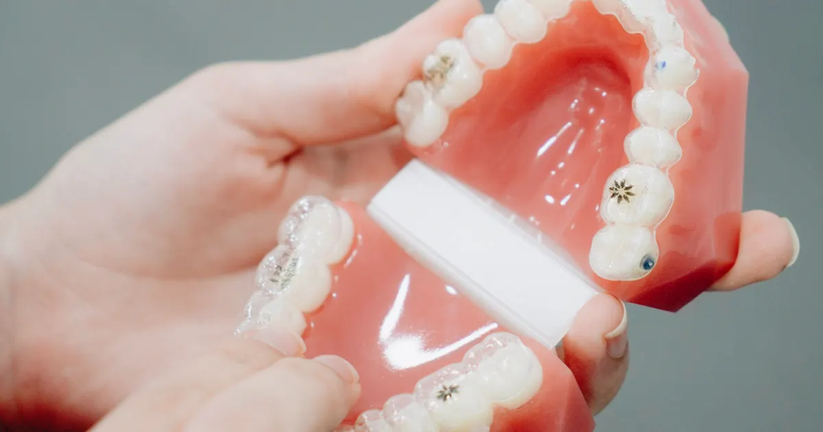
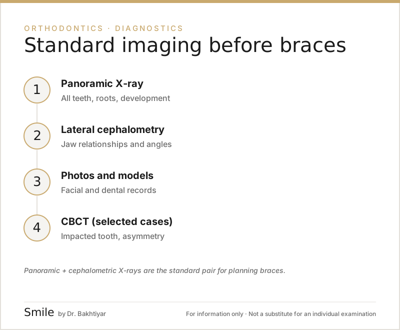
Before starting treatment with braces or clear aligners, two X-rays are taken as standard: a panoramic X-ray (OPG) and a lateral cephalometric X-ray. The panoramic view gives an overall picture of the teeth, the roots, any hidden teeth and the structure of the jaws; the cephalometric X-ray makes it possible to measure the position of the upper and lower jaw relative to each other and to the base of the skull, the angulation of the teeth and the direction of facial growth. CBCT (3D imaging) is not a routine examination — it is added only for specific indications (an impacted canine, suspected root resorption, orthodontic-surgical cases, asymmetry); periapical and bitewing X-rays are also taken when needed.
X-ray records are taken during the diagnostic stage of orthodontic treatment, based on clinical indications.
Why are X-rays essential before orthodontics?
A visual examination alone can only assess the outward appearance of the teeth. Most of the information needed for an orthodontic plan is not visible: the length and direction of the roots, unerupted (impacted) or hidden teeth, extra or missing teeth (hypodontia), the condition of the jawbone, the skeletal relationship between the lower and upper jaw, the TMJ, and the amount of bone between the tooth roots. A plan built without this information stays superficial: hidden pathology is missed, a skeletal problem goes undiagnosed (a good cosmetic result but a poor functional one), root resorption is not assessed from the outset, and attempts are made to move an impacted tooth by force.
For the difference between braces and clear aligner systems: orthodontics — braces and clear aligners.
1. Panoramic X-ray (OPG) — the teeth and the dental arch
The panoramic X-ray is the standard first examination in orthodontics: both jaws, all the teeth, unerupted teeth, the wisdom teeth, the TMJ and the sinuses are visible in a single image. The questions it answers in the orthodontic plan:
- Which teeth are present, which are missing, are there any extra teeth?
- Is there an unerupted or hidden tooth (for example, an upper canine), and in which direction is it positioned?
- Are the baby teeth still in place, and what is the eruption schedule of the permanent teeth?
- What is the length and direction of the roots, and is there any resorption?
- Is there inflammation or a cyst at a tooth root?
- What is the state of the wisdom teeth — could this change the treatment choice?
- How does the TMJ look overall?
The panoramic X-ray is particularly important in children and teenagers — the plan is adapted to the eruption schedule of the baby and permanent teeth. About the method itself: what is a panoramic X-ray?.
2. Lateral cephalometric X-ray — the skeletal relationship of the jaws
A cephalometric X-ray is a lateral image taken from the side (in profile); it shows the position of the facial bones and the jaws relative to one another in a standardised way. The orthodontist then carries out a cephalometric analysis on it (using methods such as Steiner, Downs, Ricketts and McNamara). The questions it answers:
- What is the skeletal class (I, II, III)? Is the upper jaw long or short; is the lower jaw set back or forward?
- Is growth vertical (a long face) or horizontal?
- What is the angulation of the incisors?
- Is the facial profile (lips, chin, nose) aesthetically balanced?
- Is orthognathic surgery needed, or will braces alone be enough?
In children, because the cephalometric X-ray shows the direction of growth, it makes early intervention with functional appliances possible in patients who have not yet reached their growth peak. We have a separate article on the method itself, positioning and analysis techniques: what is a lateral cephalometric X-ray?.
Why are both a panoramic and a cephalometric X-ray requested together?
Because the two X-rays answer completely different questions: the panoramic view covers the teeth and the dental arch (what is present, what is missing, what is hidden), while the cephalometric X-ray covers the skeletal relationship of the jaws (the relationship between the upper and lower jaw, the profile, the direction of growth). Together, they give the full picture of the dental and skeletal problem.
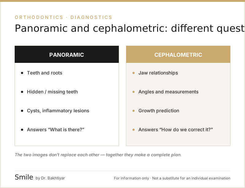
Planning with the panoramic X-ray alone — in a patient with a skeletal problem (for example, Class II — the lower jaw set back), simply aligning the teeth masks the cosmetic side of the problem; the skeletal balance is disturbed and functional problems (chewing, the TMJ, lip strain) remain. Planning with the cephalometric X-ray alone — the wisdom teeth, impacted teeth, root resorption, cysts and hypodontia are not visible; the plan looks good on paper but is risky in practice.
A practical example: in a 14-year-old teenager the upper incisors are protruding and it is difficult to close the lips. The panoramic X-ray shows that the teeth are protruding, but not why. When a cephalometric X-ray is added, it becomes clear that the lower jaw is skeletally set back (Class II), that the upper incisors are not in fact overly protrusive, and that the patient is still in the growth phase. The result: instead of simply aligning the teeth, a functional appliance (for example, a twin-block) or early orthopaedic intervention is planned. This decision is only possible by reading the two X-rays together.
The panoramic + cephalometric combination gives maximum information at the lowest dose, makes it possible to plan most orthodontic cases without needing CBCT, and is the international standard for orthodontic analysis.
3. CBCT — in which cases is it added?
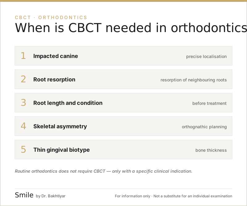
In a routine orthodontic examination (ordinary Class I cases, simple crowding) CBCT is not needed — a panoramic X-ray, a cephalometric X-ray and, if necessary, intraoral X-rays are sufficient. The 2013 position statement of the American Academy of Oral and Maxillofacial Radiology (AAOMR) recommends CBCT in orthodontics not as a routine measure, but on an individual clinical indication, with the smallest possible FOV and dose. The indications are specific:
- Impacted canine. Whether the tooth lies on the buccal (cheek) or palatal (palate) side, its effect on the neighbouring roots, the point of surgical access and the direction of traction often cannot be determined precisely with 2D methods (for example, two-angle imaging); CBCT shows these.
- Suspected root resorption. An impacted canine can resorb the root of the lateral incisor; early resorption that is not visible in 2D, the degree of damage and whether the tooth can be kept are assessed with CBCT. It is also relevant in patients who have had previous orthodontic treatment.
- Root morphology — thin, curved roots are prone to resorption during orthodontic movement; treatment forces are adjusted accordingly, especially in adult patients.
- Orthodontic-surgical cases and skeletal asymmetry — if orthognathic surgery is planned, 3D cephalometric analysis, the position of the condyle (TMJ) and virtual surgical planning.
- Cleft palate / cleft lip — the volume of the alveolar bone defect and the plan for bone grafting.
- Mini-implants (TADs) — placing a mini-screw for tooth movement with bone support.
- A thin gum biotype and a risk of gum recession — the thickness and height of the buccal cortical bone can change the movement plan.
- Complex clear aligner plans — the feasibility of difficult root movements (torque, rotation) and the limits of the aligner; CBCT is not needed for simple clear aligner treatment.
- Additional conditions in adult orthodontics — periodontitis and bone loss, root-canal-treated teeth, proximity to the nerve in the lower jaw.
- TMJ pathology if it is clinically suspected.
Can CBCT replace 2D X-rays? In theory, both a panoramic reconstruction and a 3D cephalometric analysis can be obtained from CBCT, but its dose is higher than that of the two classic X-rays, 2D cephalometry is today the standard for orthodontic analysis, and 3D is not needed in routine cases. In children (aged 7–14) CBCT is taken only in cases of an impacted tooth, cleft palate, significant asymmetry or trauma, and with the smallest FOV. One CBCT is usually enough for the whole course of treatment; a repeat scan may be needed for traction of an impacted tooth or before orthognathic surgery. In more detail: what is CBCT (3D imaging)?, when is CBCT needed.
4. Periapical and bitewing X-rays
These small intraoral X-rays play a supporting role: tooth decay (which must be treated before braces are fitted), root canal problems, and a detailed assessment of the root of a specific tooth.
Special cases
In difficult diagnoses, the orthodontist may additionally request a frontal (PA) cephalometric X-ray (if there is asymmetry), a hand-wrist X-ray (skeletal maturity — to assess growth age) or a CBCT of the TMJ.
The order in which the X-rays are taken
- Clinical examination + intraoral and facial photographs
- Panoramic X-ray
- Lateral cephalometric X-ray (usually on the same day as the panoramic X-ray)
- If needed: CBCT
- If needed: periapical / bitewing
- Digital intraoral scan (STL model)
- Orthodontic analysis (Steiner, Ricketts, McNamara, etc.) and the plan
After these steps, the patient and the dentist decide on the treatment option together: metal or ceramic braces, clear aligners, and so on. Panoramic and cephalometric X-rays brought from elsewhere can be used if they were taken within the last 6 months and are of good quality; since standard positioning is important for the analysis, the dentist sometimes recommends retaking them.
The radiation dose of the X-rays and repeat imaging
The dose of orthodontic X-rays is very low: a panoramic X-ray is around 10–25 µSv, a cephalometric X-ray around 3–6 µSv, and CBCT (small to medium FOV) approximately 30–200 µSv — together the equivalent of a few days of natural background radiation. Over a long course of treatment (18–36 months), one or two additional panoramic X-rays may be taken to check the condition of the roots and the response to treatment, and a repeat cephalometric X-ray at the end to compare the result. About the overall dose and protection: is dental X-ray dangerous?.
The pre-braces X-ray examination package in Baku
At the Smile by Dr. Bakhtiyar clinic, the standard preparation for every orthodontic patient is: digital panoramic and lateral cephalometric X-rays (at the same visit), clinical and photographic records, a digital intraoral scan, CBCT if needed, followed by cephalometric analysis in digital software and a visual explanation of the treatment options.
FAQ — frequently asked questions
1. Can braces be fitted with only a panoramic X-ray? No, a cephalometric X-ray is also needed for an orthodontic plan. The panoramic X-ray shows the teeth, the cephalometric X-ray shows the skeletal relationships — two different sets of information.
2. Does every orthodontic patient need a CBCT? No — only if there is an indication (an impacted canine, suspected root resorption, a surgical case, asymmetry, cleft palate, a mini-implant). Under the ALARA principle, CBCT is not taken without an indication.
3. Are a cephalometric X-ray and CBCT needed for clear aligners (for example, Invisalign)? A cephalometric X-ray is standard for a professional orthodontic plan and is essential in a patient with a skeletal problem. CBCT is not needed for simple clear aligner treatment; it may be recommended when complex root movements are planned.
4. Are these X-rays dangerous for teenagers and children? The dose of orthodontic X-rays is very low; the two X-rays combined are less than a few days of natural background radiation. Early diagnosis creates the opportunity to guide the growth of the jaw. The risk of missed pathology is far greater than the risk from radiation.
5. How long are X-ray results valid for? Usually 6–12 months. If treatment has not started within this period, it is recommended that they be updated.
6. Can an X-ray I bring from elsewhere be used? If the panoramic and cephalometric X-rays were taken within the last 6 months and are of good quality — yes. However, standard positioning is important for the analysis; the dentist assesses this and sometimes recommends retaking them.
7. Is a cephalometric X-ray not enough for skeletal problems — why CBCT? In most cases it is enough. For 3D asymmetry, the position of the condyle and planning orthognathic surgery, CBCT is superior.
Next step
If you have crowded teeth, a mismatch between the lower and upper jaw, aesthetic concerns or a functional problem, the first stage towards the right orthodontic plan is an accurate X-ray examination. Book an appointment — the Smile by Dr. Bakhtiyar clinic will prepare an individual plan for you.
Related reading: orthodontics — braces and clear aligners · CBCT (3D imaging) · wisdom tooth extraction.
Author: Dr. Bakhtiyar Aliyev — dentist, head of the Smile by Dr. Bakhtiyar clinic. Reviewed by a dentist: Dr. Bakhtiyar Aliyev, last reviewed: 6 July 2026.
Note: This article is for educational purposes and does not replace a consultation with a dentist. For an accurate diagnosis and treatment, please contact Dr. Bakhtiyar.
Sources
- Clinical recommendations regarding use of cone beam computed tomography in orthodontics. Position statement by the American Academy of Oral and Maxillofacial Radiology — Oral Surgery, Oral Medicine, Oral Pathology and Oral Radiology, 2013
- Optimizing radiation safety in dentistry: Clinical recommendations and regulatory considerations — Journal of the American Dental Association, 2024
- Patient risk related to common dental radiographic examinations: the impact of 2007 International Commission on Radiological Protection recommendations regarding dose calculation — Journal of the American Dental Association, 2008
This article is for general information only and does not replace an individual diagnosis, examination or treatment. Consult a doctor for a decision about your case.