

What is a lateral cephalometric X-ray?
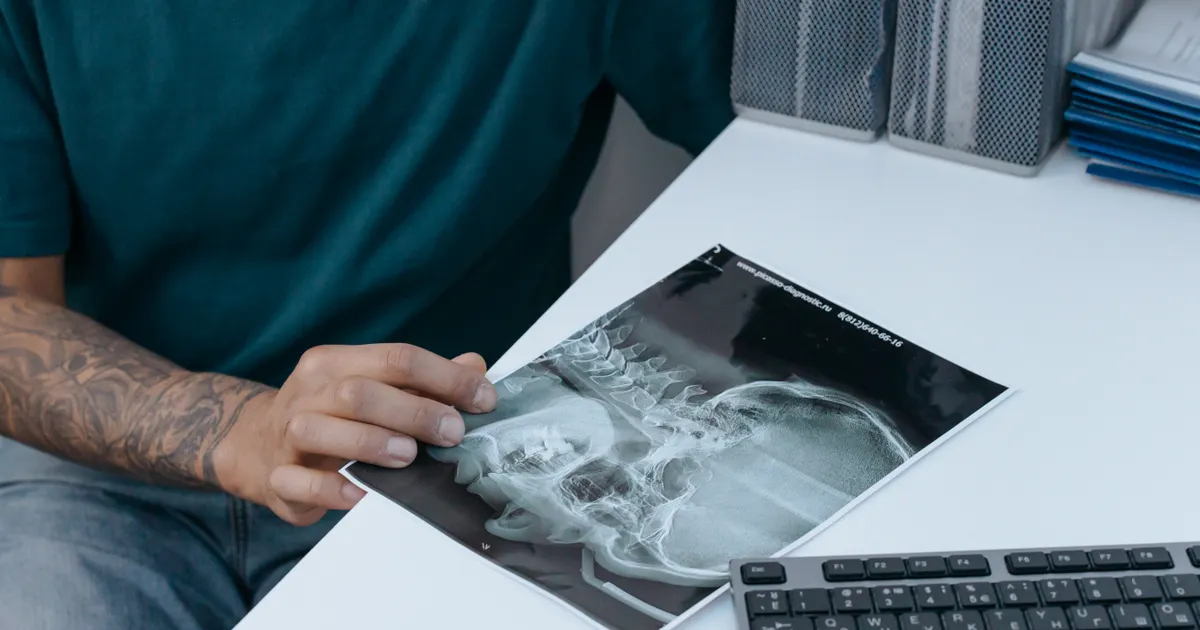
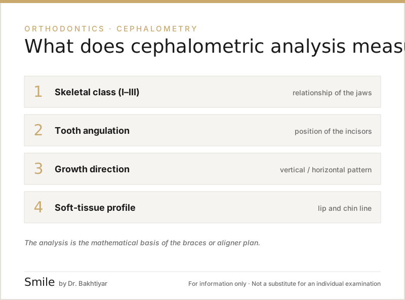
A lateral cephalometric X-ray (or lateral cephalogram) is a profile X-ray image of the head taken in a standardised position. It shows the upper and lower jaw bones, the base of the skull, the teeth and the profile outline of the soft tissues. On this image the orthodontist carries out a cephalometric analysis — measuring the position of the jaws in relation to each other and to the base of the skull, the angulation of the teeth and the direction of facial growth. It is one of the most important examinations when planning treatment with braces or clear aligners.
What does the research show? According to a systematic review (PLoS One, 2013), high-level evidence on which records are truly mandatory for orthodontic diagnosis is limited, and the study by Nijkamp and colleagues (2008) showed that in many routine cases cephalometric analysis did not change the treatment plan — which is why cephalometry is not obligatory for every patient, but is taken when indicated, such as in skeletal discrepancies, growth assessment, extraction decisions and orthognathic surgery. Variability between clinicians in identifying landmark points has also been documented, so the analysis is interpreted together with the clinical examination and study models. Related: Orthodontic services · Which X-rays are needed for braces? · What is a panoramic X-ray?.
The difference between a cephalometric X-ray and a panoramic X-ray
The two examinations are often confused, but they have different purposes:
| Feature | Panoramic X-ray | Cephalometric X-ray |
|---|---|---|
| View | from the front | from the side (profile) |
| What it shows | teeth, dental arch, TMJ | skeletal position of the jaws, profile |
| Measurement | general assessment | angle and distance measurements |
| Usually used for | initial examination, implantology, orthodontics | orthodontic planning, surgical orthodontics |
More about why a panoramic X-ray is also needed for orthodontics: which X-rays are needed for braces?.
How is a cephalometric X-ray taken?
The procedure is standardised — this is essential so that images taken in different clinics can be compared. The standard procedure:
- The patient steps up to the machine and the head is positioned in a special holder (cephalostat).
- The ear points (external auditory meatus) are held in place with fixed metal locators.
- The Frankfort horizontal plane (FH) is positioned parallel to the floor.
- The lips are kept relaxed and the teeth in maximum contact.
- The X-ray beam passes through the profile and reaches the detector.
The exposure takes just 1–2 seconds. It is painless and requires no preparation.
What is cephalometric analysis?
A cephalometric X-ray is not simply a picture — it is a diagnostic tool on which mathematical analysis is performed. The orthodontist marks reference points on the image and then calculates the angles and distances between these points.
The most commonly used reference points:
- Sella (S): the centre of the sella turcica in the skull
- Nasion (N): the centre of the frontonasal suture
- Point A: the deepest point of the cortical concavity at the front of the upper jaw
- Point B: the deepest point of the cortical concavity at the front of the lower jaw
- Menton (Me): the lowest symphyseal point of the lower jaw
- Gonion (Go): the point at the angle of the lower jaw
- ANS / PNS: the anterior/posterior nasal spine of the upper jaw
Various analysis methods have been built on the lines and angles created from these points.
Popular cephalometric analysis methods
Steiner analysis
One of the most widely used. It calculates measurements such as SNA (skeletal position of the upper jaw), SNB (skeletal position of the lower jaw) and ANB (the skeletal difference between the upper and lower jaw). It is fundamental in determining Class I / II / III skeletal relationships.
Downs analysis
Used for soft tissue profile analysis and dental aesthetics.
Ricketts analysis
Widely used to predict the direction of growth. It is particularly important in children and adolescent patients.
McNamara analysis
Assesses whether the upper jaw is positioned forwards or backwards, the length of the lower jaw and facial height.
Tweed / Wits analyses
Measure various relationships between dental and skeletal structures.
In practice the orthodontist uses one of these methods or combines several of them. Modern digital programs (for example, Dolphin Imaging, WebCeph, Nemoceph) carry out the analysis automatically.
What does cephalometry determine in an orthodontic plan?
As a result of cephalometric analysis, the dentist answers the following key questions:
- Is the problem skeletal or dental? A skeletal problem is difficult to resolve with braces alone; orthognathic surgery may be needed.
- Which jaw is long or short — the upper or the lower? Class II (lower jaw set back) and Class III (lower jaw set forward) problems are diagnosed with this analysis.
- What is the angulation of the teeth? The forward or backward inclination of the incisors is analysed.
- What is the direction of growth? In child patients, a vertical (long face) or horizontal (short face) growth pattern.
- Profile analysis. The soft tissues play a key role in facial aesthetics.
Without this information it is still possible simply to align the teeth, but the result may not be optimal functionally and aesthetically.
Frontal (posteroanterior) cephalometry
Standard cephalometry is taken laterally (from the side). However, in some cases a frontal cephalometric X-ray (posteroanterior, PA) is also taken. This is taken with the patient facing forwards. Frontal cephalometry is used in assessing jaw asymmetries.
When is cephalometry needed?
- Before orthodontic treatment — a standard indication (skeletal discrepancy, growth, extraction decision)
- Orthognathic surgery planning — a standard indication
- Growth monitoring — repeated periodically in children and adolescents
- At the end of treatment — to compare the results
Cephalometry is not needed for routine dental treatment or for implants.
The difference between cephalometry and CBCT
CBCT provides a three-dimensional volumetric image and many analyses can also be carried out on it (3D cephalometry). However:
- The dose from CBCT is several times higher than that of classic cephalometry
- Conventional 2D cephalometry is still fundamental in international orthodontic analysis standards
- In simple cases 3D is not needed — 2D is sufficient
A CBCT scan is chosen for surgical orthodontics, impacted canines and complex asymmetries. More: CBCT (3D imaging).
Digital cephalometry
Today most clinics carry out cephalometry digitally. The image is stored in DICOM or JPEG format and analysed within a software program. The advantages of digital analysis:
- faster and more accurate landmark marking (with AI support)
- easy comparison with previous analyses
- assessment of the treatment response with visual overlay (superimposition)
- easy explanation of the summaries and reports to the patient
Radiation safety
A cephalometric X-ray is a very low-dose examination — on average 5–7 μSv. This is equal to or less than the natural background radiation received on a flight to the seaside. It is also considered safe in children, particularly when taken with a proper indication.
In pregnant women, planned orthodontic X-rays are usually postponed until after the birth — unless there is an urgent indication.
Can a patient understand the cephalometry results themselves?
Usually not — the results become meaningful with the dentist's explanation. However, it is important for the patient to find answers to the key questions:
- What is my class? — I, II or III?
- Is my problem mainly dental or skeletal?
- Is surgery needed?
- Is the growth phase working in my favour? (for children)
In a good clinic, the results of the cephalometric analysis are also explained to the patient visually.
The cephalometric X-ray process in Baku
At the Smile by Dr. Bakhtiyar clinic, digital cephalometry is taken for every orthodontic patient. The analysis is carried out using WebCeph and other modern programs, and the results are explained to the patient visually. This makes it easier both to choose the treatment plan and to compare the results at the end of treatment.
FAQ — frequently asked questions
1. Is a cephalometric X-ray painful? No, it is completely painless and takes 1–2 seconds.
2. Is the radiation safe? Yes — the dose is very low. In children and adolescents too, the risk is considered very low when it is taken with a proper indication.
3. Can I see how my face will look using cephalometry? Digital programs can give an approximate picture of how the facial profile will change after treatment. This is called digital treatment planning.
4. Does my child need cephalometry? If an orthodontic problem is suspected — yes. Because the child is in a growth phase, the analysis and the timing of any intervention are important.
5. Is cephalometric analysis automatic or done by hand? Digital programs offer automatic analysis, but the dentist checks and corrects it. In practice a semi-automatic approach is the standard.
Next step
If you would like a digital and accurate cephalometric analysis for your orthodontic plan, book an appointment. At the Smile by Dr. Bakhtiyar clinic, panoramic and cephalometric X-rays are taken digitally and the results are explained to you clearly.
Related reading: which X-rays are needed for braces? · orthodontics — braces and clear aligners · CBCT (3D imaging).
Author: Dr. Bakhtiyar Aliyev — dentist, head of the Smile by Dr. Bakhtiyar clinic. Reviewed by a dentist: Dr. Bakhtiyar Aliyev, last reviewed: 6 July 2026.
Note: This article is for educational purposes and does not replace a consultation with a dentist. For an accurate diagnosis and treatment, please contact Dr. Bakhtiyar.
Sources
- Records needed for orthodontic diagnosis and treatment planning: a systematic review — PLoS One, 2013
- The influence of cephalometrics on orthodontic treatment planning — European Journal of Orthodontics, 2008
- Cephalometric landmark variability among orthodontists and dentomaxillofacial radiologists: a comparative study — Imaging Science in Dentistry, 2015
This article is for general information only and does not replace an individual diagnosis, examination or treatment. Consult a doctor for a decision about your case.