

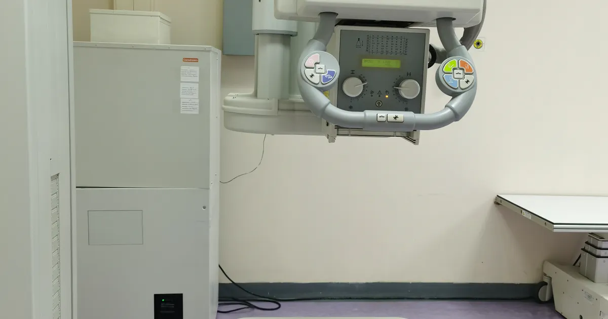
CBCT (Cone Beam Computed Tomography) is a 3D imaging method used in dental radiology. Its dose depends on the FOV: small FOV 30–100 µSv, medium FOV 100–200 µSv, large FOV 200–1000 µSv. It is higher than a panoramic X-ray (10–24 µSv), but 5–20 times lower than a conventional head CT scan (1500–2500 µSv). When taken on clinical indication, its contribution to cancer risk is statistically negligible, but repeating it without a reason is unacceptable (ALARA).
The clinic's indication-based dose approach (ALADA, smallest possible FOV) is explained on the 3D diagnostics page.
The most frequently asked question about CBCT is: Is the radiation high? The answer is not a short one — because high is a relative concept. Compared with a panoramic X-ray, yes, it is higher in any case. Compared with a conventional medical CT scan, it is much lower. In this article I answer the question together with its context.
This article is about CBCT only. For general dental X-ray safety, a dose table covering all types of X-ray and the question of how many times you can have one taken, see: Is dental X-ray dangerous?. For a comparison of 2D and 3D: The difference between panoramic X-ray and CBCT.
What exactly is the dose of a CBCT scan?
The CBCT dose depends on the field of view (FOV — Field Of View), the device model and the protocol. Typical values are:
| CBCT type | FOV | Typical dose |
|---|---|---|
| Small FOV (around a single tooth, 4×4 cm) | ~4×4 cm | 30–100 µSv |
| Medium FOV (one jaw) | ~8×8 cm | 100–200 µSv |
| Large FOV (both jaws + sinus) | ~15×15 cm or more | 200–1000 µSv |
| Low Dose CBCT | variable | considerably lower than the standard protocol for the same FOV |
These ranges may look wide — the reason is the large difference between devices. A meta-analysis published in 2015 (Ludlow et al., Dentomaxillofacial Radiology), based on data pooled from 20 studies, reports the following average effective doses for adults with standard protocols: small FOV 84 µSv, medium FOV 177 µSv, large FOV 212 µSv; while for individual devices, values between 46 and 1073 µSv were recorded for large FOV. In other words, the dose of a single CBCT scan can be 2–40 times that of a panoramic X-ray — depending on the FOV and the device. On how the FOV is chosen: What is FOV and how is it chosen?.
What counts as high? — Context matters
The comparisons below give a practical answer to the question of high:
- One small FOV CBCT (50 µSv) ≈ one week of natural background radiation (world average 8–10 µSv/day).
- One medium FOV CBCT (150 µSv) ≈ two weeks of natural background or 1.5 chest X-rays.
- One large FOV CBCT (500 µSv) ≈ two months of natural background or one mammogram.
- One conventional head CT (2000 µSv) ≈ ten months of natural background or four large FOV CBCT scans.
So the CBCT dose is significantly lower than that of a conventional CT scan, but it is the highest within the dental X-ray family. This gradation is the basis of clinical decision-making: for each problem, the method with the lowest dose that still provides sufficient information is chosen.
Why does the CBCT dose vary so much?
Under the same name of CBCT, the dose can differ by 10–20 times. The reason lies in four CBCT-specific factors:
- FOV (the area scanned) — the larger the area, the greater the irradiated volume and the dose; a small FOV covers a few teeth, a large FOV the entire face and jaws (What is FOV and how is it chosen?).
- Voxel size (resolution) — high resolution such as 75–100 µm requires more frames and a longer exposure; for implant planning 200–300 µm is enough and the dose is lower.
- Protocol — number of frames, mA, kVp, rotation time (a full 360° or a half rotation); Low Dose modes reduce these.
- Device — detector sensitivity and the manufacturer's standard settings; the meta-analysis recorded large differences between devices for the same FOV.
This is why the correct answer to the question of what the dose of a CBCT scan is always begins with another question: which FOV, which resolution, which protocol?
Why is the CBCT dose higher than that of a panoramic X-ray?
The reason is technical. A panoramic X-ray produces a single flat image — with successive frames taken along the face. CBCT, on the other hand, collects projections from hundreds of different angles in order to build a volumetric model. Each projection delivers a small portion of radiation, and together they add up to a higher dose.
But this extra dose brings very valuable information — 3D anatomical accuracy, bone width, the true 3D distance to the nerve canal, pathology inside the sinus. These are things a panoramic X-ray cannot provide.
What is Low Dose CBCT?
Low Dose CBCT is a scanning mode of 3D tomography specifically designed to use less radiation. The device parameters are optimised according to the clinical indication; as a result, the dose for the same FOV drops considerably below the standard protocol, while diagnostic quality is preserved for most clinical needs.
How is the dose reduced?
During a classic CBCT scan, the device collects several hundred X-ray frames (projections) as it rotates around the head; each frame is a separate exposure. Low Dose mode lightens this process in several ways:
- Reducing the number of frames — the software fills in the gaps mathematically;
- Reducing the mA (milliamperes) and the exposure time — for example, an 8-second rotation instead of 14 seconds;
- Choosing a small FOV — only the necessary region is scanned;
- Matching the voxel size to the need — for implant planning a voxel of 200–300 µm is usually enough, 100 µm is not required;
- Fast rotation — a shorter exposure;
- Iterative / artificial-intelligence-based reconstruction — a high-quality volume is built from fewer frames.

Who is the Low Dose protocol suitable for?
- Children and adolescents — the dose must be kept to a minimum in a growing body;
- When a repeat CBCT is needed — for example, a second scan during orthodontic treatment or implant follow-up;
- Initial planning before an implant — a medium voxel is enough for bone height and width;
- General orthodontic assessment — an overall picture of unerupted teeth and the jaw structures;
- Patients with a high dose history — for example, those under oncological follow-up;
- Initial bone assessment of the TMJ.
When is it not enough?
In cases requiring micro-detail, the standard (high-resolution) protocol is preferred: searching for a root fracture or a small apical lesion, additional canal anatomy, orthognathic surgery planning, suspicion of cancer, and narrow-field plans requiring sub-millimetre accuracy. The mode is chosen not by the patient but by the referring dentist or the radiologist: the clinical question, age, dose history and the expected diagnostic need are assessed together. Both modes are available on the same device through the software, and Low Dose does not involve any extra cost.
Clinical risk assessment
For one small FOV CBCT (50 µSv), the contribution to cancer risk calculated under the linear no-threshold model is approximately 1 in 500,000 — that is, statistically orders of magnitude smaller than everyday lifetime risks (travelling by car, ordinary household accidents).
For one medium FOV CBCT (150 µSv) this figure is around 1 in 150,000, and for a large FOV CBCT (500 µSv) around 1 in 50,000. In international publications, these risks are regarded as statistically immeasurable.
The scientific position on CBCT's contribution to cancer risk is therefore as follows:
- It is not zero, but it is very low.
- When taken on clinical indication, the benefit-risk balance is positive.
- Repeating it without a reason is not acceptable under the ALARA principle.
In which clinical situations does CBCT add value?
The higher dose of CBCT is clinically justified in the following situations:
- Implant planning — bone width, nerve canal, sinus.
- Complex wisdom tooth surgery — to reduce the risk of nerve injury.
- Sinus lift planning.
- Root canal treatment — detecting microcracks and hidden canals.
- Orthodontic surgery — complex anatomy.
- Surgical planning for cysts and tumours.
- Trauma — following the fracture line precisely.
- TMJ — assessment of the bone structure of the temporomandibular joint.
In all of these scenarios a panoramic X-ray alone is not sufficient, so the CBCT dose is justified by the clinical outcome.
What does too many CBCT scans mean for one patient?
In ordinary clinical life, the lifetime number of CBCT scans for one patient is usually:
- 1–3 CBCT scans over a lifetime — with an average dental history.
- 5–7 CBCT scans — in a patient who has had implant and orthodontic treatment.
- 10+ CBCT scans — rare, with a very complex medical history.
Even with 10 CBCT scans (in the large FOV version) the total dose is about 5 mSv — two to three times that of a single head CT scan. This is not considered statistically significant for lifetime radiation accumulation.
CBCT in children — caution and indication
Children are more sensitive to radiation. In children, CBCT is taken only on a strong clinical indication:
- Planning for a hidden permanent tooth.
- Surgical planning for an impacted tooth (canine, incisor).
- Cleft palate or a syndromic anatomical need.
- Trauma.
In these cases a small FOV + Low Dose protocol is applied. According to the meta-analysis data, the average dose for a small FOV with a standard protocol in children is about 103 µSv (Ludlow et al., 2015); Low Dose mode brings this figure down further. More details: Are dental X-rays safe for children?.
CBCT during pregnancy
In pregnant patients, CBCT is not taken electively — all elective scans are postponed until after the birth. If there is an acute need (jaw trauma, a growing cyst), the dentist assesses the risk-benefit balance, and in very rare cases the scan is done with special protection. More on this: Can you have an X-ray taken during pregnancy?.
Steps patients themselves can take to reduce the dose
- Ask the clinic about a Low Dose protocol.
- Ask whether the clinic will take the CBCT with a small FOV.
- If you have a previous CBCT — always show it to your dentist (a repeat scan may not be needed).
- If the reason is not clear — ask your dentist to explain the clinical indication.
- Keep the DICOM file yourself — other clinics may then not need to repeat the scan.
Is it high? — The answer
- Compared with a panoramic X-ray: Yes, it is 2–40 times higher depending on the FOV.
- Compared with a conventional head CT scan: No, it is significantly lower (5–20 times).
- In terms of clinical purpose: When there is a clinical indication, the accuracy this dose provides makes it clinically justified.
- For a scan without a reason: No, the let us just take one anyway approach does not suit CBCT.
It is normal for the answer to this question to be neither yes nor no — because a medical decision is always read together with the clinical indication.
What to look for when having a CBCT in Baku?
- The device being digital and up to date.
- The availability of small FOV and Low Dose protocols.
- The existence of a paediatric protocol.
- Results provided in DICOM format.
- The scan being read by a specialist with certified radiology training or a specialisation in dental radiology.
You can see these details on our CBCT service page.
Frequently asked questions (FAQ)
Is the CBCT dose higher than that of a panoramic X-ray?
Yes, it is 2–40 times higher depending on the FOV. But it is still a dose 5–20 times lower than a conventional head CT.
Does CBCT cause cancer?
The contribution of one CBCT scan to cancer risk is statistically very small — it is a dose equivalent to one day of natural background radiation. When taken on clinical indication, the benefit outweighs the risk.
What is Low Dose CBCT?
It is a low-dose protocol — the number of frames, the mA, the exposure time and the voxel size are reduced in line with the clinical need; the dose for the same FOV is considerably lower than with the standard protocol. It is used in children, for repeat scans and for initial planning.
Is Low Dose CBCT less accurate?
It is accurate enough for most clinical indications (implant planning, orthodontic assessment, follow-up). For root fractures, small apical lesions and cases requiring micro-detail, the standard high-resolution protocol is chosen.
Does Low Dose CBCT cost a different price?
Usually not. The mode is changed through the software on the same device; no extra cost is involved.
What is FOV and how does it affect the dose?
FOV (Field Of View) is the size of the area being imaged. The larger it is, the higher the dose. Whenever possible, a small FOV is chosen.
Can CBCT be taken every year?
On clinical indication. For active implant follow-up or surgical preparation, 1–2 CBCT scans a year is considered normal; a preventive annual scan is not recommended.
About the author
Dr. Bakhtiyar Aliyev — dentist, clinical director of the Smile by Dr. Bakhtiyar clinic. This article has been medically reviewed by Dr. Bakhtiyar.
This article is for general information purposes and does not replace individual medical advice.
Book an appointment
For a modern, low-dose CBCT scan in Baku, you are welcome to contact our clinic. Book an appointment online or get in touch with us.
Sources
- Effective dose of dental CBCT — a meta analysis of published data and additional data for nine CBCT units — Dentomaxillofacial Radiology, 2015
- Optimizing radiation safety in dentistry: Clinical recommendations and regulatory considerations — Journal of the American Dental Association, 2024
- ICRP Publication 103: The 2007 Recommendations of the International Commission on Radiological Protection — International Commission on Radiological Protection, 2007
This article is for general information only and does not replace an individual diagnosis, examination or treatment. Consult a doctor for a decision about your case.