

What is axiography: how is the movement of the jaw joint recorded?
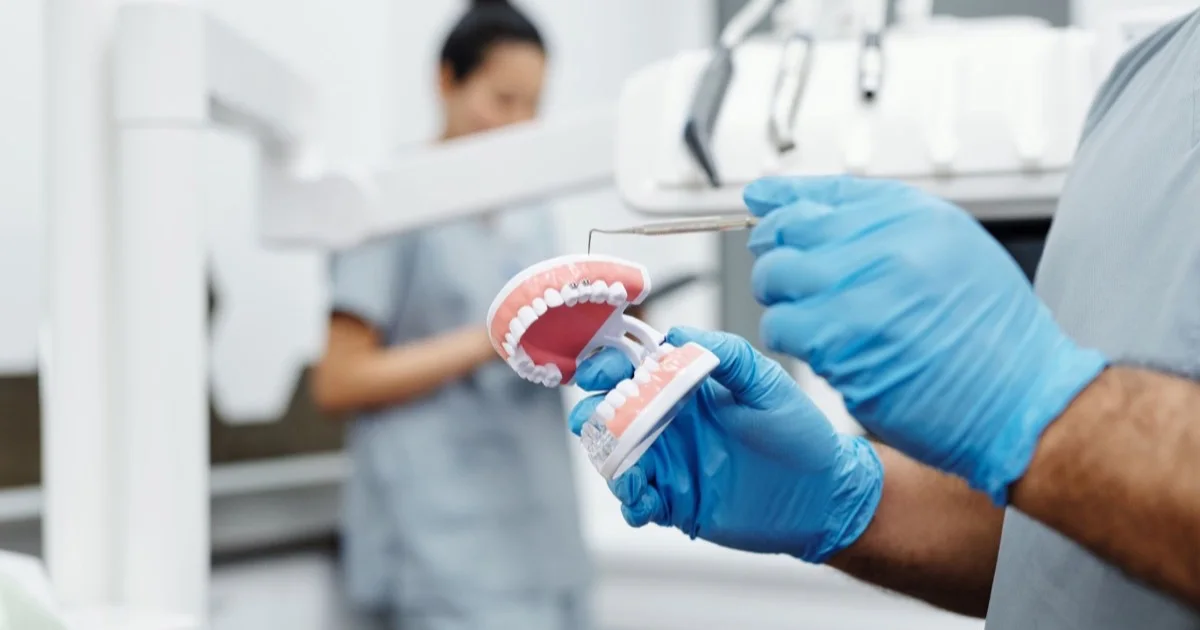
Axiography (also called condylography) is a functional examination that records the path travelled by the head of the lower jaw (the condyle) when you open your mouth and move it forwards and sideways. Sensors attached to the head and to the lower jaw trace the movement on a computer as a curve. X-rays and MRI show the structure of the joint; axiography shows how the joint works. It involves no radiation, is painless and takes 20–30 minutes. It does not make a diagnosis on its own — together with a clinical examination it is used to assess joint problems and to plan large prosthetic work.
Key points
- Axiography measures movement, not structure: the length, shape and inclination of the condylar path and the coordination of the two sides are recorded.
- Main uses: functional evidence when disc displacement is suspected, and individual programming of the articulator for full-mouth prostheses.
- In studies, standardised assessment gives high agreement between dentists.
- There is no radiation; there are practically no contraindications.
- Not every TMD patient needs it — the indication is determined by the dentist.
How does the jaw joint move?
When you open your mouth, the condyle first rotates inside the fossa (rotation) and then slides forwards and downwards, along the bony ridge called the articular eminence (translation). Together these two movements create a curved path. The right and left condyles should move at the same time and at a similar speed. The articular disc (the cartilage cushion between the condyle and the bone) accompanies the condyle along this path.
When there is a problem, this path changes: if the disc has slipped forwards, the condyle catches until it reaches it and then jumps (a click); if there is inflammation, the path shortens and asymmetry appears between the two sides. Axiography is what makes these changes visible.
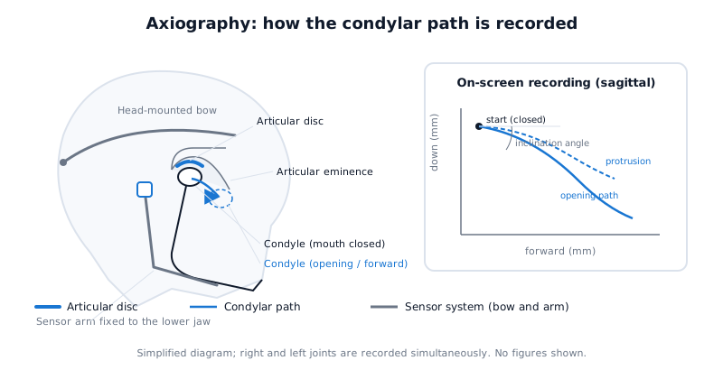 The sensors record the path the condyle travels from the joint fossa along the articular eminence; the recording appears on the screen as a curve.
How is the examination carried out?
- Preparation. A light bow (the upper part) is placed on the head; a small device that sticks to the lower teeth temporarily and painlessly is attached, together with the sensor arm connected to it.
- Calibration. The device finds the joint axis of the condyle — the hinge axis. This is why the name of the examination comes from the word axio- (axis).
- Movements. Following the dentist's instructions, you open and close your mouth and move your jaw forwards (protrusion) and to both sides (laterotrusion). Each movement is repeated several times.
- Recording. The computer draws the path for each condyle as a curve in two planes — from the side (sagittal) and from above (horizontal).
- Analysis. The dentist assesses the length and shape of the path, the stability of the start and end points, the synchrony of the two sides and the speed of movement.
Modern devices are electronic (for example, Cadiax) or based on ultrasound; some systems work with optical tracking. The principle is the same — the movement is recorded and converted into numbers.
What is measured and what do the results tell us?
- Sagittal condylar path inclination — the angle at which the condyle descends as it moves forwards. This figure is used for the individual programming of the articulator (a laboratory device that simulates jaw movement); the chewing surface of the prosthesis is shaped accordingly.
- The length and shape of the path — a short path indicates limited movement, while a broken or stepped curve gives information about the disc catching.
- The stability of the start and end points — whether the condyle returns to the same place on closing.
- Coordination of the two sides — whether the right and left condyles move at the same time and whether there is a difference in speed.
In a study published in 2019 (34 patients, 34 healthy controls), in people with internal derangement of the joint (disc displacement) the condylar path on the working side was longer during lateral movement, while the condyle on the opposite side was faster. In other words, a disc problem changes the pattern of movement, and this change shows up in the recording.
Does axiography make a diagnosis?
Partly. In a study published in 2022, computerised axiography was compared with MRI (the reference examination for the condition of the disc): in detecting disc displacement, sensitivity was around 85%, specificity 95% and overall accuracy 88% (in the same patients, against MRI; the measure was detection of disc displacement). At the same time, the authors stress that without a clinical examination axiography has no diagnostic meaning in itself; it is a complementary tool.
There is evidence on reliability as well. In a controlled, blinded study published in 2023, four trained dentists assessed the computer recordings of 20 patients using standard criteria; overall agreement was high (Fleiss kappa 0.88 — almost perfect agreement; the measure was interpretation of the recordings). The lowest agreement was in assessing condylar coordination, which led to the criteria being refined. The conclusion: when read with a standard protocol, axiography is not a subjective eyeball estimate but a repeatable measurement.
Who is it recommended for?
Axiography is not a routine examination. It is usually useful in the following situations:
- Suspected disc displacement — clicking, catching, deviation to one side on opening; as functional evidence before MRI or alongside it.
- Full-mouth prosthetic and rehabilitation planning — for individual programming of the articulator when multiple crowns, an implant-supported prosthesis or a change in bite height is planned. This may be part of a full mouth rehabilitation plan.
- Orthodontic or orthognathic planning — to document the initial functional situation in cases where the jaw position will be changed.
- Monitoring splint treatment — to compare the pattern of movement before and after occlusal splint treatment.
For simple muscle pain, short-term jaw fatigue or clenching that is purely stress-related, axiography is usually not needed — a clinical examination is enough. You can read about when symptoms are serious in the article symptoms of TMJ problems.
The information it provides for bite and prosthetic planning
The most widely used side of axiography is not diagnosis but planning. When several crowns, an implant-supported bridge or a full-mouth prosthesis is being made, the laboratory sets the teeth up on an articulator. The articulator is a model of your jaw: it has two joint mechanisms and these are set at certain angles. When these angles are set with average figures, the prosthesis is made for an average person; when they are set from a recording of your own joint, it is made for you. Axiography provides exactly these figures.
Condylar path inclination (sagittal condylar guidance)
As the condyle moves forwards, it descends along the slope of the articular eminence. The angle of this slope — how many degrees it makes with the horizontal plane — is called the condylar path inclination. When the angle is steep, the back teeth separate quickly as the jaw moves forwards; when it is shallow, the teeth stay closer together and the likelihood of collisions on the chewing surface increases. This angle therefore decides how high and steep the cusps of the back teeth (the peaks on the chewing surface) can be.
There are several ways of measuring the inclination: a wax protrusive bite record, a panoramic or lateral cephalometric X-ray, CBCT and axiography. A systematic review published in the Journal of Prosthetic Dentistry in 2024 (24 studies, dentate adults) showed that the methods give different figures: the difference between the wax record and the panoramic X-ray was up to 9 degrees; wax records came out statistically lower than pantograph records. The authors currently consider CBCT the most suitable method, and note that digital axiography may surpass the other methods because it captures the functional information in full. The practical conclusion: the method used for the measurement matters as much as the figure itself, and the laboratory needs to know it.
Bennett movement and the side shift
When you move the jaw to the right, the right condyle (the working side) rotates in place and moves slightly sideways; the left condyle (the balancing side) slides forwards, downwards and inwards. This lateral component is called the Bennett movement. It has two parts: a short, straight sideways shift that occurs at the very start of the movement (the immediate side shift) and the sloping path that continues afterwards, forming an angle with the sagittal plane (the Bennett angle).
Why are these parameters important for a prosthesis? Because during lateral movement the cusps of the back teeth have to pass by one another. When the side shift is large, the pathways between the cusps have to be opened wider and shallower; otherwise an interference arises on the balancing side during chewing — the patient feels this as one tooth touching first, and in the long run may face a cracked crown or joint and muscle discomfort. Axiography records the lateral movement separately for each condyle; the dentist transfers this to the articulator.
Programming the articulator
On a semi-adjustable articulator, two figures are usually entered: the condylar path inclination and the Bennett angle; on a fully adjustable articulator, the immediate side shift, the intercondylar distance and the curvature of the path are set as well. In a digital workflow the same parameters are entered into a virtual articulator — the CAD software — and the chewing surface of the prosthesis is checked on the computer with a movement simulation. The axiographic recording provides these figures directly; some systems transfer the recording to the articulator software as a file.
How much better are individual figures than average ones? The evidence is still cautious. In a preliminary clinical study published in 2025 (20 patients, single crowns, designed with CAD), designs with no parameters, with average parameters and with fully individual parameters were compared: in the design without parameters the patients' discomfort scores were higher, and both of the two crowns that cracked came from this group; no statistical difference was found between average and individual parameters (short follow-up; the outcomes were occlusal contact area and patient comfort). In simple terms: for a single crown, average figures are often enough; the value of an individual recording increases when joint movement differs noticeably from the average figures, when the two sides are asymmetrical, or when the whole chewing surface is being rebuilt.
Its effect on the design of the chewing surface
These figures translate into the shape of the teeth as follows:
- Cusp height and inclination. A steep condylar path allows steeper cusps; a shallow path requires the cusps to be lower and softer.
- The direction of the grooves and pathways. The Bennett angle and the side shift determine in which direction and how widely the grooves on the chewing surface of the back teeth are opened.
- Harmony with anterior guidance. The inner surface of the front teeth separates the back teeth as the jaw moves forwards; this anterior guidance must be built in harmony with the condylar path, otherwise either the back teeth do not separate or the front teeth take excessive load.
- When the bite height changes. When the bite height is increased, the condyle takes up a new position in the fossa; the movement recording helps to check in advance how the teeth will meet in this new position.
We have explained separately how the subject of the bite is linked to the joint in the article dental occlusion and the TMJ.
The limits of this section
- Axiography does not track the condyle itself but a reference point calculated from the device attached to the teeth; the measured inclination may differ by a few degrees depending on the method.
- The lateral movement parameters (Bennett angle, immediate side shift) have a smaller amplitude than the sagittal inclination and are more sensitive to the quality of the recording.
- Long-term comparative evidence on the effect of individual parameters on the lifespan of a prosthesis is limited; the available studies have short follow-up and small samples.
- However precisely the articulator is set, checking in the mouth and small corrections (occlusal adjustment) are usually still needed.
The difference from X-rays, CBCT and MRI
| Examination | What it shows | Radiation | Relation to axiography |
|---|---|---|---|
| Axiography | Path of movement, inclination, coordination | None | Function |
| CBCT | Bone structure: shape of the condyle, arthritic changes | Yes (low dose) | Structure; read together with function |
| MRI | Soft tissue: position of the disc, inflammation | None | Reference for the disc |
| Panoramic X-ray | General overview | Yes (low dose) | Initial screening |
We have an article on the place of CBCT in joint problems — TMJ problems on CBCT; and we have compared separately which examination is chosen for which question in MRI, CBCT and axiography for TMJ pain. In short: structure and function do not replace one another; the dentist looks at both when needed.
Limitations
- Because the sensors are attached to the skin and to the teeth, the point measured is not the condyle itself but a reference point close to it; recordings are interpreted on this basis.
- The inclination of lateral (transverse) movements is measured less reliably than the sagittal inclination.
- The result depends on the dentist's analysis and on the calibration; a trained specialist is required.
- With very limited mouth opening or acute pain, a complete recording may not be possible.
- The relationship between the bite and joint problems is complex; axiography does not prove this relationship, it only documents movement. We have explained this subject separately in the article dental occlusion and the TMJ.
In our clinic the functional assessment is carried out by an orthodontist-gnathologist; the workflow is set out on the TMJ and gnathology page.
Frequently asked questions
Is axiography painful and how long does it take? It is painless. The device attached to the teeth is temporary and leaves no trace. Preparation and recording together usually take 20–30 minutes.
Does axiography replace MRI? No. MRI shows the position of the disc directly; axiography records the pattern of movement. When a disc problem is suspected, the two examinations complement each other; sometimes a clinical examination plus axiography reduces the need for MRI, and sometimes it is the other way round.
Is axiography needed for a single crown too? Usually not. For a single crown, the average parameters of the articulator and checking in the mouth are often enough. Axiography becomes valuable with multiple crowns, when the bite height is changed, or when joint movement is asymmetrical.
Is axiography carried out in children? It is technically possible, but in a developing jaw the indications are narrow and individual; it is done at the dentist's discretion.
If the result is normal, does that mean I have no joint problem? No. Pain of muscular origin or early inflammation may not change the condylar path. A normal recording reduces the likelihood of a structural disorder, but the diagnosis is made together with the clinical picture.
Conclusion
Axiography is a radiation-free functional examination that makes the movement of the jaw joint visible and measurable. It provides additional evidence when disc displacement is suspected; in large prosthetic and orthodontic plans it individualises the articulator with figures such as the condylar path inclination and the Bennett movement, and it guides the design of the chewing surface. It does not make a diagnosis on its own; it is read together with the clinical examination and, when needed, imaging. Whether you need this examination can be clarified at a consultation with an orthodontist-gnathologist.
Sources
- Condylar motion analysis: a controlled, blinded clinical study on the interindividual reproducibility of standardized evaluation of computer-recorded condylar movements — Scientific Reports, 2023 (Ahlers et al.)
- Diagnostic use of computerized axiography in TMJ disc displacements — Experimental and Therapeutic Medicine, 2022 (Talmaceanu et al.)
- Computerized ultrasonic axiographic evaluation of condylar movement in patients with internal derangement of the temporomandibular joint — The Angle Orthodontist, 2019 (Choi et al.)
- Sagittal condylar guidance angle measurement methods: A systematic review — The Journal of Prosthetic Dentistry, 2024 (Lassmann et al.)
- Occlusal Analysis of Algorithm-designed Restoration Using Different Virtual Articulator Parameters: A Preliminary Study — International Dental Journal, 2025 (Guo et al.)
This article is for general information only and does not replace an individual diagnosis, examination or treatment. Consult a doctor for a decision about your case.