

The Difference Between CBCT and a Conventional CT Scan
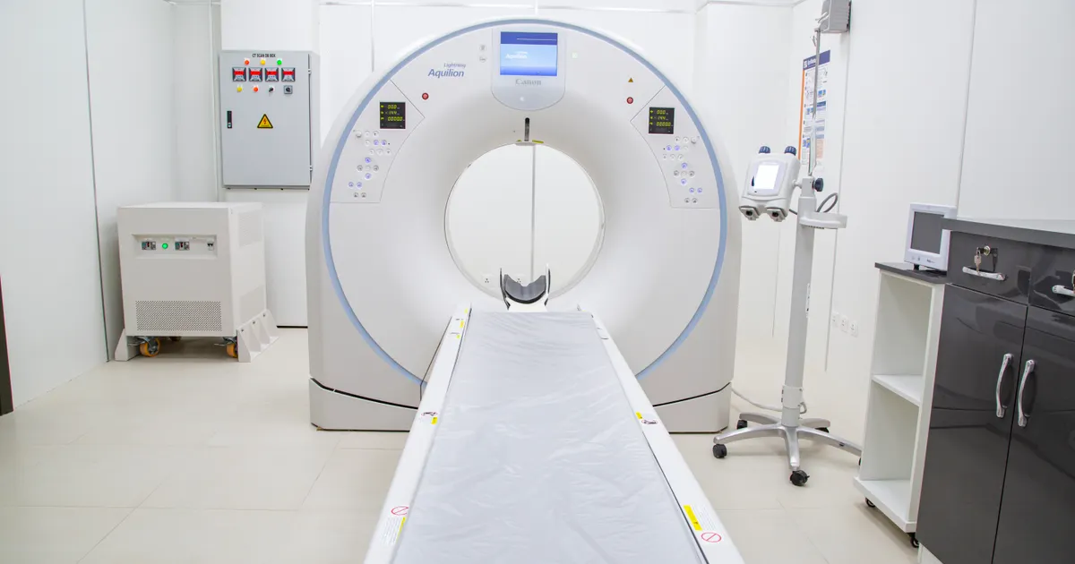
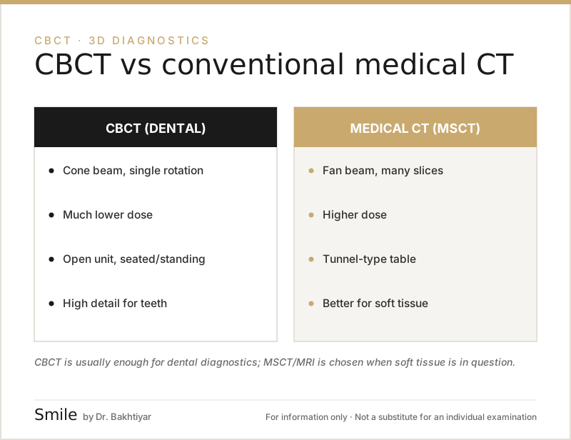
Both CBCT and a conventional CT (MSCT) show the jaw in 3D, but they work with different technologies. CBCT scans in a single rotation using a cone-shaped beam, the dose is usually considerably lower (several times lower, depending on the field of view and the mode), and it gives ideal accuracy for teeth, bone and the nerve canal. A conventional CT, on the other hand, is superior for soft tissues and tumours, but involves a higher dose.
What does the research show? CBCT collects a volume in a single rotation using a cone beam and a flat panel detector, whereas MSCT takes spiral slices with a fan beam — this gives CBCT high spatial resolution (bone, teeth, canals) and gives MSCT superior soft tissue contrast and Hounsfield density measurement (Dental Clinics of North America, 2008). In terms of dose: in a meta-analysis, the effective dose of dental CBCT averaged ~131 μSv for a large FOV, ~88 μSv for a medium FOV and ~34 μSv for a small FOV, while a medical head CT is usually in the 1–2 mSv range — so the difference is usually large, but it narrows with large fields of view and high-quality modes, which is why the claim that CBCT always means less radiation is not universally true. AAOMR/AAE position papers recommend choosing between the two methods according to the clinical question. Related: 3D diagnostics (CBCT) service · What is CBCT? · Is CBCT radiation high?.
The choice between these two technologies is not arbitrary: each has its own place. In this article we explain the differences, the advantages, the doses and which one is taken in which situation, through a dentist's eyes.
Quick comparison table
| Feature | CBCT (dental 3D) | Conventional CT (MSCT) |
|---|---|---|
| Beam shape | Cone (cone beam) | Fan beam, spiral |
| Scan time | 15–25 seconds | 30 seconds – several minutes |
| Voxel shape | Isotropic (equal sided) | Anisotropic (different layers) |
| Bone imaging | Very high accuracy | High accuracy |
| Soft tissue | Poor | Very good (with contrast agent) |
| Dose (µSv) | ~40–200 | ~1000–2000+ |
| Machine size | Compact, in the clinic | Large, in a hospital |
| Price | Relatively low | Considerably higher |
| Main indication | Teeth, jaw, sinus, TMJ | Whole body: tumours, trauma, vessels |
The key difference in how they work
The fundamental difference between the two lies in the shape of the beam and in the detector.
Conventional CT (Multi-Slice CT / MSCT): A thin, fan-shaped beam rotates in a spiral around the jaw and records the jaw as hundreds of parallel slices. The computer then combines these slices to create a 3D image. This method is optimised to show soft tissues as well.
CBCT (Cone Beam CT): A wide, cone-shaped beam covers the entire exposure area at once. The detector is a flat panel and captures several hundred frames. All the 3D data is collected in just one rotation (~15–25 seconds). This method is optimised for teeth, bone and nerve canals.
As a result, CBCT gives more accurate information about bone and teeth, while a conventional CT gives richer information about soft tissues.
Radiation dose — the most important difference
The difference in dose is very large with a small field of view, and smaller with wide FOV and high-quality modes; this is why the choice is always made according to the clinical question.
- Dental CBCT (small FOV, single region): ~40–100 µSv
- CBCT (large FOV, full jaw + sinus): ~100–200 µSv
- Standard medical head CT: ~1500–2000 µSv
- MSCT for the sinuses: ~600–1000 µSv
In other words, for typical implant planning, CBCT means about as much dose as a short to medium-haul flight; taking a conventional CT for the same purpose means unnecessary radiation. For this reason, international guidelines (SEDENTEXCT, EU) list CBCT as the first choice for dental indications.
We have written in more detail about why the dose matters and about the overall safety of CBCT in our article What is CBCT (3D tomography).
Accuracy and the voxel difference
The main technical advantage of CBCT is its isotropic voxel. A voxel is a unit of measurement similar to a pixel, but in 3D.
- In CBCT the voxel has equal sides (e.g. 0.2 × 0.2 × 0.2 mm). This means you can take measurements with the same high accuracy in any plane.
- In MSCT the voxel is usually an elongated cuboid (e.g. 0.5 × 0.5 × 1.0 mm). This is accurate along the main scanning direction, but looks thick in other planes.
This difference is very important for implant and orthodontic planning: when sub-millimetre measurement is required, CBCT is at least equal and often superior.
Contrast agent — a major difference
- MSCT can work with an intravenous contrast agent (iodine-based). This shows vascular structures, inflammation and tumours very clearly.
- Contrast is not used in CBCT. Its purpose is different: accuracy in hard tissues.
That is why, when a tumour or a vascular problem is suspected, the patient is referred to a radiology centre — CBCT cannot show these well enough.
Which one should be chosen and when?
CBCT is recommended for:
- Dental implant planning (bone height, width, density)
- Difficult root canal treatment (extra canals, root curvature)
- Wisdom tooth removal (proximity of the tooth to the nerve)
- Orthodontic treatment planning (impacted teeth, root position)
- The temporomandibular joint (TMJ) — assessment of the bone structure
- Toothache of sinus origin — the relationship between upper jaw teeth and the sinus
- Trauma to the teeth and jaw (fractures)
A conventional CT (MSCT) is recommended for:
- Soft tissue tumours and lymph nodes
- Vascular pathologies (contrast-enhanced CT angiography)
- Serious trauma and multiple facial fractures
- Detailed assessment of large cysts and tumours (spread into soft tissue)
- Oncological staging
See our articles on which situations make CBCT essential, When is CBCT absolutely necessary, and on which conditions can be seen, Which conditions are visible on CBCT.
The FOV (field of view) difference
CBCT has a freely selectable FOV — the dentist scans only the region that is needed. This makes it possible to keep the dose low:
- Small FOV (4×4 cm) — a single tooth, root canal treatment
- Medium FOV (8×8 cm) — a jaw segment, implant
- Large FOV (13×15 cm or more) — the full jaw, sinuses, TMJ
With MSCT, the scan area is usually large and non-selective — software parameters are used to reduce the dose.
Speed and comfort
CBCT takes 15–25 seconds in a single rotation; MSCT of the jaw can last from 30 seconds to 1 minute, and the spiral movement together with the large number of slices creates a risk of artefacts. A short scan time is important for children and anxious patients — the less movement, the clearer the image.
Price and availability
CBCT is available in most dental clinics today, and its price in Baku is at an affordable level. A conventional CT, on the other hand, is taken in hospitals and large radiology centres and costs considerably more. For a purely dental indication, CBCT is superior both medically and economically.
FAQ — the most frequently asked questions
1. Are CBCT and MSCT the same thing?
No. Both provide 3D, but the technology, the dose and the area of use are different. For dental indications the first choice is CBCT; for soft tissue and vessels, MSCT.
2. My dentist recommends CBCT — could a CT at the hospital not be used instead?
Usually not. A medical CT involves a higher dose and does not provide the accuracy needed for teeth. The right thing is to have a CBCT taken at the clinic.
3. Which method has the lower dose?
In terms of dose, CBCT is lower. However, if the indication concerns soft tissue or vessels, MSCT is required — and in that case the dose is medically justified.
4. Is it worth taking a CBCT instead of a panoramic + 2D X-ray?
Only if there is a clinical indication. For a routine dental check-up, a panoramic and 2D X-ray are enough. For implants, root canals, orthodontics and so on, CBCT is superior.
5. Does metal show up on CBCT?
Metal implants and crowns sometimes create artefacts, but with special filters (MAR) most artefacts are reduced. Diagnostic information is not lost.
Conclusion and appointment
CBCT and conventional CT do not replace one another — they are complementary methods. The dentist makes the choice by looking at which question needs to be answered. For the vast majority of dental problems, CBCT — low dose, fast and accurate — is the first choice.
If an implant, root canal treatment or jaw surgery is being planned for you and CBCT has been recommended, this is an important step towards making the treatment safe and precise.
👉 Book an appointment for CBCT and our other services at our clinic in Baku.
Author: Dr. Bakhtiyar Aliyev — dentist, chief dentist of the clinic. This article has been reviewed by a dentist.
Sources
- What is cone-beam CT and how does it work? — Dental Clinics of North America, 2008
- Effective dose of dental CBCT — a meta analysis of published data and additional data for nine CBCT units — Dentomaxillofacial Radiology, 2015
- AAE and AAOMR Joint Position Statement: Use of Cone Beam Computed Tomography in Endodontics — 2015 Update — Oral Surgery, Oral Medicine, Oral Pathology and Oral Radiology, 2015 (AAE/AAOMR)
- Position statement of the American Academy of Oral and Maxillofacial Radiology on selection criteria for the use of radiology in dental implantology with emphasis on cone beam computed tomography — Oral Surgery, Oral Medicine, Oral Pathology and Oral Radiology, 2012 (AAOMR)
This article is for general information only and does not replace an individual diagnosis, examination or treatment. Consult a doctor for a decision about your case.