

How is bone measured for a dental implant? Height, width and density on CBCT
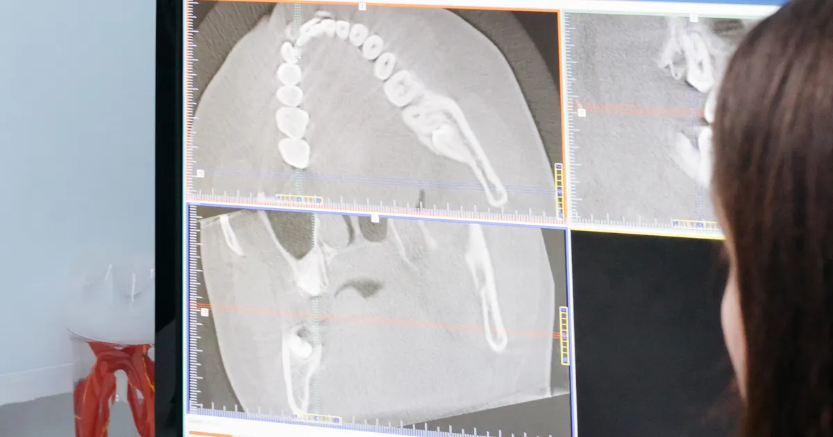
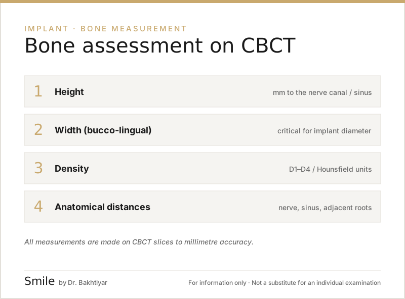
The thickness of the jawbone — both its width and its height — is today measured most accurately with CBCT (3D cone-beam computed tomography). Using special software, the dentist creates cross-sectional slices of the CBCT scan and takes measurements in millimetres. The height, width and density of the alveolar bone are assessed in this way — and this is the foundation of implant, bone grafting and orthodontic planning. This article is about measurement and 3D assessment; the treatment options available when the dimensions are not sufficient are covered in a separate article: When there is not enough bone for an implant.
Why is bone thickness measured?
Bone thickness is a decisive parameter in implantology and orthodontics. There are two main dimensions:
- Height (vertical) — the distance from the alveolar crest to the nerve canal or the sinus floor.
- Width (horizontal, buccolingual) — the thickness of the bone between the cheek and tongue sides.
The generally accepted minimums for a standard implant:
- Height: at least 10 mm (keeping a safe distance from the nerve and the sinus)
- Width: at least 6 mm (so that 1.5 mm of bone remains on each side around the implant)
If these dimensions are smaller, either short/narrow implants are used or bone grafting is planned. If the measurement is not accurate, the implant may fail, the edge of the bone may resorb, and the nerve may even be damaged. For this reason, measuring ‘by eye’ or ‘approximately’ is not acceptable.
Methods used to measure bone thickness
Although several methods have existed historically, the main methods used in the clinic today are the following.
1. CBCT — the gold standard of three-dimensional measurement
CBCT is the most accurate measuring tool in implantology today. The machine rotates once around the patient’s head and produces a volumetric (3D) image of the jaw. The dentist loads this image into special software (for example, Blue Sky Plan, coDiagnostiX, Romexis) and can create slices in any plane:
- axial slice — allows the jaw to be viewed from above downwards
- coronal slice — a slice running from the front towards the back
- sagittal slice — a slice from the side
- panoramic reconstruction — the 3D version of the 2D panoramic image
The most important of all is the cross-sectional slice. This is the slice that shows both the height and the width at the same time at the site of each tooth. With a millimetre ruler, the dentist can take a measurement at any point.
More about CBCT: CBCT (3D diagnostics).
2. Panoramic X-ray — only an approximate height
A panoramic X-ray is a two-dimensional examination. It gives no information about the width of the bone, and it shows the height with distortion and magnification errors. A panoramic X-ray is useful for an initial assessment, but on its own it is not enough for implant planning.
3. Clinical palpation
During surgery, the surgeon can measure the width of the bone with a finger or with a special bone caliper. This is an additional method of confirmation, but it is not used for planning in advance — because it requires additional surgical access.
The bone measurement process on CBCT, step by step
After the CBCT scan, the measurement steps proceed as follows:
- Loading the image into the software. The DICOM file is opened in the planning software.
- Setting the reference plane. The guiding axis (along the alveolar arch) is drawn.
- Selecting the cross-sectional slices. In the area of tooth loss, slices are created at 1–2 mm intervals.
- Measuring the height. Measured in millimetres from the alveolar crest to the upper border of the nerve canal or to the sinus floor.
- Measuring the width. The distance between the buccal (cheek) and lingual/palatal (tongue/palate) cortical plates.
- Assessing the density. The bone type is determined using Hounsfield units (HU) or digital indices: D1 (the densest) to D4 (the softest).
- Adding a safety margin. A distance of at least 1.5–2 mm is kept from the nerve canal and the sinus.
Why is bone density important?
It is not only thickness but also the quality (density) of the bone that affects the outcome. Soft bone (type D4, mainly in the posterior region of the upper jaw) makes the initial stability of the implant more difficult. Dense bone (D1) is found in the anterior region of the lower jaw and often provides high stability. Knowing this information, the dentist:
- selects the diameter and length of the implant
- modifies the surgical technique (for example, reducing the osteotomy speed in soft bone)
- plans the loading time (immediate, early or delayed)
Standard minimum dimensions — table
| Structure | Minimum height | Minimum width |
|---|---|---|
| Standard implant (Ø 4 mm) | 10 mm | 6 mm |
| Narrow implant (Ø 3–3.5 mm) | 10 mm | 5 mm |
| Short implant | 6–8 mm | 6 mm |
| Zygomatic implant (special case) | different | different |
These dimensions are a general guide, and every case is assessed individually by the dentist.
Options when there is not enough bone
If the CBCT shows that the bone is insufficient, there is no need to panic. Modern dentistry offers several reliable ways forward:
- Bone grafting (augmentation): bone is built up with autogenous (your own bone), allogeneic or synthetic materials
- Sinus lifting: in the posterior region of the upper jaw, the sinus floor is lifted and bone is placed beneath it
- Short implants: for bone with limited height
- Tilted implants / All-on-4: placed at an angle in the area where bone is available
More details: bone grafting and sinus lifting · when there is not enough bone for an implant.
Bone loss in areas where teeth have been extracted
After a tooth is extracted, the alveolar bone resorbs most rapidly during the first 3–6 months. Within one year, horizontal loss can reach 25%, and within four years 40–60% (Schropp et al.). For this reason, CBCT measurements often do not remain reliable for a long time. If an implant is being planned, a fresh measurement is recommended.
More details: tooth loss and jawbone loss.
Bone thickness in orthodontics
The orthodontist also pays attention to bone thickness — particularly:
- when planning tooth movement (if the alveolar bone is thin, the risk of root resorption increases)
- for the placement of orthodontic mini-implants (TAD — Temporary Anchorage Device)
- for surgical orthodontic planning in adult patients
How does the bone measurement process work in Baku?
At the Smile by Dr. Bakhtiyar clinic, bone measurement is a 3-step process:
- Clinical examination and panoramic X-ray — for a general assessment.
- CBCT scan — for the area where an implant or bone grafting is planned.
- Digital planning — virtual implant placement in the software and, if necessary, the preparation of a surgical guide.
All measurements are also provided to the patient together with the DICOM file.
FAQ — frequently asked questions
1. Can an ordinary panoramic X-ray show bone thickness? No, a panoramic X-ray does not show the width of the bone and shows the height with distortion. CBCT is needed for an accurate measurement.
2. Is the X-ray for bone measurement painful? No, it is completely painless. A CBCT scan takes 15–40 seconds and the patient simply needs to stand still.
3. If there turns out to be little bone, does that mean an implant is impossible? No. Most cases can be resolved with bone grafting, sinus lifting or alternative implant strategies. The dentist makes the decision after reviewing the CBCT result.
4. Do bone dimensions change over time? Yes. After tooth loss, bone resorption continues. For this reason, older CBCT scans often need to be updated (usually after 6–12 months).
5. Does bone thickness decrease with age? Generally — yes. In older patients, mineral density and the height of the alveolar bone may decrease, particularly in women with osteoporosis.
Next step
If you are planning an implant or orthodontic treatment, have your bone thickness measured precisely with a professional CBCT assessment at the Smile by Dr. Bakhtiyar clinic. Book an appointment — Dr. Bakhtiyar will prepare the treatment plan that best suits your measurements.
Related reading: CBCT (3D diagnostics) · when there is not enough bone for an implant · tooth loss and jawbone loss.
Author: Dr. Bakhtiyar Aliyev — dentist, head of the Smile by Dr. Bakhtiyar clinic. Reviewed by a doctor: Dr. Bakhtiyar Aliyev, date of last review: 6 July 2026.
Note: This article is for educational purposes and does not replace a consultation with a dentist. For an accurate diagnosis and treatment, please contact Dr. Bakhtiyar.
This article is for general information only and does not replace an individual diagnosis, examination or treatment. Consult a doctor for a decision about your case.