

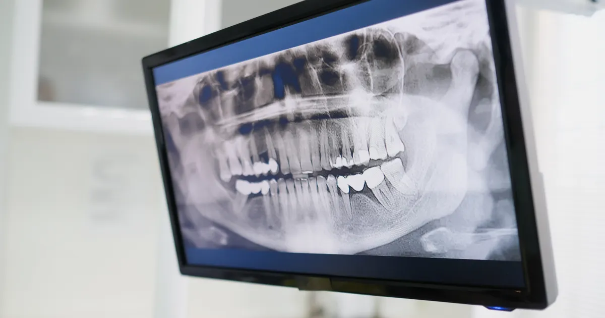
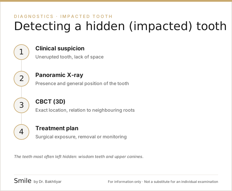
A hidden tooth (impacted tooth) is a tooth that has not been able to erupt fully or partially even though it is due to come through, and has remained under the jawbone or the gum. The most common hidden teeth are the wisdom teeth (third molars) and the upper canines. Hidden teeth are first suspected during a clinical examination, then detected with a panoramic X-ray, while their exact position and relationship with neighbouring structures are determined with CBCT (3D tomography).
What is a hidden tooth and why does it happen?
A tooth normally forms inside the alveolar bone and, at a certain age, erupts into the jaw and takes its place. Sometimes:
- there is not enough room (the dental arch is small)
- the tooth tilts in the wrong direction
- the tooth meets another tooth on its eruption path (for example a baby tooth)
- a cyst or pathological tissue blocks the eruption path
For these reasons the tooth stays in the jaw and becomes hidden (impacted). Sometimes it erupts only partially and remains halfway — this happens especially with wisdom teeth and causes pericoronitis (inflammation around the tooth).
Which teeth are most often hidden?
- Wisdom teeth (3rd molars) — the teeth that most often remain hidden. They cannot erupt because of a lack of space in the lower and upper jaw. More on this: wisdom tooth extraction.
- Upper canines — the second most commonly impacted teeth after wisdom teeth. They cause orthodontic and aesthetic problems.
- Lower premolars — in rare cases.
- Incisors — when a baby tooth is lost prematurely or after trauma.
When does the suspicion of a hidden tooth arise?
Clinically, the dentist sees that a tooth is still not visible even though its eruption age has passed. Standard eruption ages:
| Tooth | Usual eruption age |
|---|---|
| Lower incisors | 6–8 years |
| Upper incisors | 7–9 years |
| Upper canines | 11–13 years |
| Premolars | 10–12 years |
| Second molars | 11–13 years |
| Wisdom teeth (3rd molars) | 17–25 years |
If these ages have passed and a tooth still has not come through, a hidden tooth is suspected. Sometimes a child's parents come in worried that a new tooth is not coming through.
Steps in detection
1. Clinical examination
The dentist checks inside the mouth and identifies the gap in the dental row. The gum is checked by hand — a hidden tooth can sometimes be felt beneath the gum.
2. Panoramic X-ray (OPG)
A panoramic X-ray is the tool for the initial detection of hidden teeth. The whole jaw is visible — which tooth is hidden, its current position and the bone around it are immediately apparent.
What a panoramic X-ray shows:
- the presence of a hidden tooth
- its general direction (vertical, horizontal, tilted)
- the stage of root formation (it may not yet be fully mature)
- a cyst or pathological increase in size around it
- in the case of a wisdom tooth — its approximate proximity to the nerve canal in the lower jaw
However, a panoramic X-ray is two-dimensional. It cannot fully show whether the tooth is tilted towards the palate or towards the lip. That is why three-dimensional imaging is needed in complex cases.
3. CBCT — precise three-dimensional localisation
CBCT is invaluable when deciding whether a hidden tooth should be removed or guided orthodontically into its correct position.
What CBCT shows:
- whether the tooth is on the palatal/labial side
- whether there is pressure on and resorption of the neighbouring roots
- the exact distance to the nerve canal in the lower jaw
- proximity to the sinus floor in the upper jaw
- a cyst or odontogenic pathology
- the thickness of the buccal / palatal bone
Especially important: upper canines often tilt towards the palate (palatal side). This is not fully visible on a panoramic X-ray — CBCT shows it precisely. This information is decisive when planning orthodontic traction (pulling the tooth into place).
More about CBCT: CBCT (3D diagnostics).
4. Periapical X-ray — Clark's SLOB technique
In some cases two periapical X-rays are taken from different angles and compared — this allows an initial estimate, using Clark's SLOB (Same Lingual, Opposite Buccal) rule, of whether the canine lies on the lingual or the buccal side. It is a classic technique, but it falls behind CBCT in terms of accuracy when CBCT is available.
Problems caused by a hidden tooth
An undetected hidden tooth causes problems over time:
- root resorption of the neighbouring tooth — the most serious. A hidden canine can press on and dissolve the root of the neighbouring incisor.
- pericoronitis — inflammation around a partially erupted wisdom tooth
- cyst formation (dentigerous cyst) — a fluid-filled cyst around the tooth
- gaps in the dental arch and orthodontic problems
- infection and swelling
- impaired chewing function
For this reason, once a hidden tooth is detected, the treatment decision should not be postponed.
Treatment options
The main treatment choices after detection:
1. Monitoring
If the tooth is not harming health and is not moving, monitoring is chosen in some cases. Usually for wisdom teeth that are not yet fully formed at a young age.
2. Surgical removal (extraction)
The most commonly chosen treatment for wisdom teeth and for some impacted teeth that cause problems. Before surgical removal, CBCT is important for assessing proximity to the nerve canal.
3. Surgical exposure + orthodontic traction
Especially for upper canines. The tooth is surgically exposed, an orthodontic bracket is bonded to it and it is pulled into place month by month. This process can take many months, but it makes it possible to preserve the tooth.
More about orthodontic treatment options: orthodontics — braces and clear aligners.
4. Removal of a cyst / pathological tissue
If there is a cyst around the hidden tooth, it is removed during the same operation and sent for histological analysis.
Hidden teeth in children
In children, the main hidden tooth problem is the premature loss of baby teeth and, as a result, the blocking of the eruption path of the permanent tooth. For this reason, timely treatment is important. More on this: tooth decay in baby teeth.
If there are specific clinical indications (for example, if a canine has not erupted despite the child being over 13), a panoramic X-ray is recommended.
Hidden teeth and the orthodontic plan
Detecting hidden teeth in advance is essential for orthodontic treatment:
- the possibility of pulling the hidden tooth into place is assessed
- surgical exposure is planned if needed
- wisdom teeth are discussed beforehand (extraction during or after orthodontic treatment)
- anchorage is set up with mini-implants
Orthodontic treatment started without these steps can become difficult and the results can be put at risk.
The hidden tooth detection process in Baku
At the Smile by Dr. Bakhtiyar clinic, for a patient with a suspected hidden tooth:
- Clinical examination + intraoral photographs
- Panoramic X-ray — initial detection
- CBCT if needed — 3D localisation
- Orthodontic and/or surgical consultation
- Explanation of treatment options and a plan
FAQ — frequently asked questions
1. Can a hidden tooth be felt by hand? In some cases the bulge of the tooth can be felt over the gum — but in most cases nothing is felt. That is why a clinical examination + X-ray are important.
2. Does a hidden tooth always have to be removed? No. The decision depends on the type of tooth, its position, the pathology around it and the orthodontic plan. Sometimes monitoring is chosen, sometimes removal, sometimes pulling it into place.
3. Does a hidden tooth cause pain? Not always. There is pain when wisdom teeth cause pericoronitis; other hidden teeth often give no symptoms at all and are detected on an X-ray.
4. My child's baby tooth has stayed in place for a long time — is this a sign of a hidden tooth? Sometimes yes. A baby tooth remaining in the jaw for a long time can be a sign that the permanent tooth beneath it is impacted. A panoramic X-ray confirms this.
5. Which X-ray is best for a hidden tooth? A panoramic X-ray for initial detection, CBCT for precise localisation. CBCT plays a particularly big role for upper canines.
Next step
If you or your child have a suspected hidden tooth, modern imaging methods are needed for precise detection. Book an appointment — at the Smile by Dr. Bakhtiyar clinic a full assessment is carried out with a panoramic X-ray and, if needed, CBCT.
Related reading: wisdom tooth extraction · CBCT (3D diagnostics) · orthodontics — braces and clear aligners.
Author: Dr. Bakhtiyar Aliyev — dentist, head of the Smile by Dr. Bakhtiyar clinic. Reviewed by a dentist: Dr. Bakhtiyar Aliyev, last review date: 6 July 2026.
Note: This article is for educational purposes and does not replace a consultation with a dentist. For an accurate diagnosis and treatment, please contact Dr. Bakhtiyar.
This article is for general information only and does not replace an individual diagnosis, examination or treatment. Consult a doctor for a decision about your case.