

What is peri-implantitis and why does inflammation develop around an implant?
Peri-implantitis is inflammation of the tissues around an implant together with the gradual loss of the bone that supports it. Its early stage is peri-implant mucositis: the inflammation is limited to the gum, no bone has been lost yet, and with timely treatment the situation can be reversed. The main cause is bacterial plaque that builds up on the surfaces of the implant and the crown — the body responds to it with inflammation. The diagnosis is not made by looking alone, but by gentle probing of the gum and an X-ray.
Key points
- Peri-implant mucositis — inflammation of the gum only, without bone loss. Peri-implantitis — inflammation + ongoing bone loss.
- Both are linked to plaque (biofilm). A crown shape that is difficult to clean, cement left under the gum and missed check-ups all speed the process up.
- Gentle probing and an intraoral X-ray are needed for the diagnosis. That is why the first (baseline) measurements should be recorded when the prosthesis is fitted.
- Inflammation caught early is simple to manage; once bone loss has begun, treatment is stepwise and often surgical.
- Previous periodontitis, smoking, poor hygiene and diabetes are risk factors.
Mucositis and peri-implantitis: what is the difference?
The gum around a healthy implant looks like the gum around a healthy tooth: there is no redness or swelling. When the dentist probes with light force (sources cite approximately 0.2 N), there is no bleeding. On the X-ray, after the natural bone remodelling of the healing period, the bone level stays stable.
In mucositis there are signs of inflammation — first and foremost bleeding on probing, sometimes redness, swelling, even pus — but there is no bone loss. In peri-implantitis, alongside the inflammation, the supporting bone is progressively lost. Whether or not there is bleeding on probing is decisive for the diagnosis.
In simple terms: mucositis is a reversible condition that stays at gum level. Peri-implantitis is structural damage that does not fully restore the bone that has been lost. Not every case of mucositis turns into peri-implantitis, but mucositis is considered its precursor and a risk factor. According to the sources, peri-implantitis can begin as early as within the first three years after the prosthesis is fitted, and it sometimes progresses rapidly.
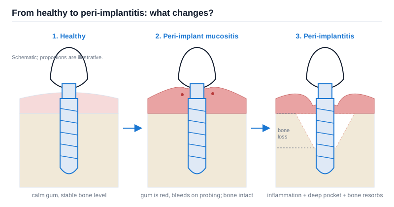 Simplified cross-sections: the inflammation starts in the gum first; once the bone level drops, the condition is already called peri-implantitis. The proportions are schematic.
Why does inflammation develop around an implant?
Bacterial plaque and the body's response
Peri-implant diseases are inflammatory complications that arise around implants integrated with bone. The mechanism is simple: microbial plaque builds up at the neck of the implant and around the abutment and the crown, the body responds to it with inflammation, and the balance is disrupted. Experimental studies in humans have shown that there is a direct cause-and-effect relationship between plaque accumulation and the onset of inflammation.
That is why the idea that “an implant is an artificial material, so it cannot become inflamed” is wrong. The inflammation takes place not in the implant itself, but in the living tissues that surround it.
Local factors that make the process easier
- A crown shape that is difficult to clean. If the edge of the crown leaves no access for a toothbrush, an interdental brush or a water flosser, plaque stays there.
- A deeply seated crown margin. An experimental study has shown that when an implant is placed deep and the crown margin sits well below the gum, a deep pocket forms and it becomes harder for the inflammation to heal completely.
- Cement left under the gum. With cement-retained crowns, excess cement left under the gum becomes a nest for plaque.
- Lack of follow-up. When you do not come for check-ups for years, early changes go unnoticed.
Patient-related factors
The risk factors listed include a previous diagnosis of periodontitis (gum disease), poor plaque control, smoking and diabetes. The effect of these factors on implants is a separate topic — you can read our article on smoking, diabetes and other risk factors. Our article on periodontitis is also useful when it comes to gum disease itself.
How does peri-implantitis differ from periodontitis?
Both are plaque-related inflammation, but the structure of the tissues is different. In a natural tooth, the healthy connective tissue fibres between the site of inflammation and the bone act as a kind of barrier. Around an implant there is no such fibre attachment.
Studies carried out in animal models show that a peri-implantitis lesion can spread as far as the bone marrow spaces and, because there is no healthy fibre layer in front of it, advances unhindered. The practical consequence: in some patients the process moves faster than expected. That is why early diagnosis, timely treatment and regular monitoring after treatment are especially important.
Which signs should you watch for?
In the early stage there may be few complaints. An implant has no nerve of its own, so pain often comes late. Watch for:
- bleeding around the implant when brushing or spontaneously;
- redness, swelling or tenderness of the gum;
- pus when you press on it, a persistent bad taste or odour;
- gum recession, a feeling that the crown is getting longer;
- the crown or the screw becoming loose and, rarely, the implant becoming mobile.
The last of these may already be a late picture. We have discussed when you need to be seen urgently in more detail in our article on the signs of implant failure.
How is the diagnosis made?
The diagnosis rests on three things.
1. Probing. With a thin instrument, the dentist gently measures the depth of the pocket between the gum and the implant and notes whether there is bleeding. This should be done at every check-up visit.
2. X-ray. An intraoral X-ray is needed to confirm a diagnosis of peri-implantitis. If no earlier image and measurements are available, the sources state that three conditions must be present together for the diagnosis: bleeding on probing, a pocket of 6 mm or deeper, and a bone level 3 mm or more below the top of the intraosseous part of the implant.
3. Baseline records. It is recommended that the first X-ray and probing measurements be kept after the prosthesis is fitted. In later years, any change is compared precisely against these. 3D imaging (CBCT) can give additional information about the bone on the cheek and tongue sides, but it is not recommended as a routine monitoring method.
How is it treated?
Treatment is stepwise and depends on the severity of the condition.
Removing the cause. First, the conditions are created for plaque to be cleaned away without obstruction: the shape of the crown is corrected, excess cement is removed, and you are taught an individual hygiene technique. Without this step, the remaining stages do not give lasting results.
Non-surgical stage. The implant and abutment surfaces are cleaned with professional instruments and, if necessary, antiseptic agents are added. The situation is then reassessed. In mucositis this stage is often enough.
Surgical stage. If the inflammation continues and there is bone loss, the gum is opened and the area is cleaned; depending on the shape of the defect, methods that smooth or rebuild the bone are discussed. If bone loss has advanced a great deal, removing the implant also remains an option.
One important note: a Cochrane systematic review states that there is no reliable evidence allowing us to determine which method is more effective in treating peri-implantitis. This does not mean that the methods do not work — it simply means that the “best method” has not yet been proven. That is why the plan is drawn up individually, and the outcome is always monitored with repeat examinations. The clinical guideline of the European Federation of Periodontology (EFP) also recommends a stepwise, team-based approach.
Can it be prevented?
No treatment comes with a full guarantee, but there are real ways to reduce the risk:
- Daily hygiene — cleaning around the implant every day with an interdental brush, super floss or a water flosser.
- Professional care — cleaning and check-ups scheduled at individually set intervals; the detailed routine is in our article on long-term care after an implant.
- Early intervention — not waiting once you see bleeding; at the mucositis stage the situation is far more manageable.
- Managing risk factors — stopping smoking, keeping diabetes under control, treating periodontitis both before and after the implant.
- Proper planning — the 3D position of the implant and a crown shape that can be cleaned; this is part of the dental implant placement plan.
Frequently asked questions
Can inflammation around an implant be painless? Yes. In the early stage the main sign is not pain, but bleeding when brushing or on probing. So even if you have no complaints, do not skip your scheduled check-ups.
Does peri-implantitis go away on its own? No. As long as the plaque is not removed, the inflammation continues. Mucositis can be managed with professional cleaning and proper home care; once bone loss has begun, professional treatment is needed.
Does a diagnosis of peri-implantitis mean the implant definitely has to be removed? No. The decision depends on the extent of the bone loss, the shape of the defect, how firm the implant is and how easy it is to keep clean. Removal is discussed only when restoration is not possible.
Does lost bone come back? With some defect shapes, regenerative methods aim to restore it partially, but this is not possible in every case and the result is individual. The realistic goal is often to stop the process and stabilise the situation.
I have had gum disease on my natural teeth. Can I have an implant? Yes, but the periodontitis must be treated first, and then a close monitoring schedule must be set up — because a history of periodontitis is considered a risk factor.
Conclusion
Peri-implantitis is not an event that suddenly “ruins” an implant. It usually develops over a long period, starting with inflammation of the gum. That is why the most effective strategy is to catch the early stage: daily hygiene, scheduled check-ups, baseline X-rays and probing records. To assess your individual situation, the condition of your existing implants and the right monitoring interval can be discussed during a consultation.
Sources
- Peri-implant mucositis and peri-implantitis: key features and differences — British Dental Journal, 2024
- Guideline on treatment of peri-implant diseases (EFP S3 level clinical practice guideline) — European Federation of Periodontology, 2023
- Peri-Implant Diseases — American Academy of Periodontology
- Interventions for replacing missing teeth: treatment of peri-implantitis — Cochrane Database of Systematic Reviews, 2012
This article is for general information only and does not replace an individual diagnosis, examination or treatment. Consult a doctor for a decision about your case.