

What does a cyst look like on an X-ray?
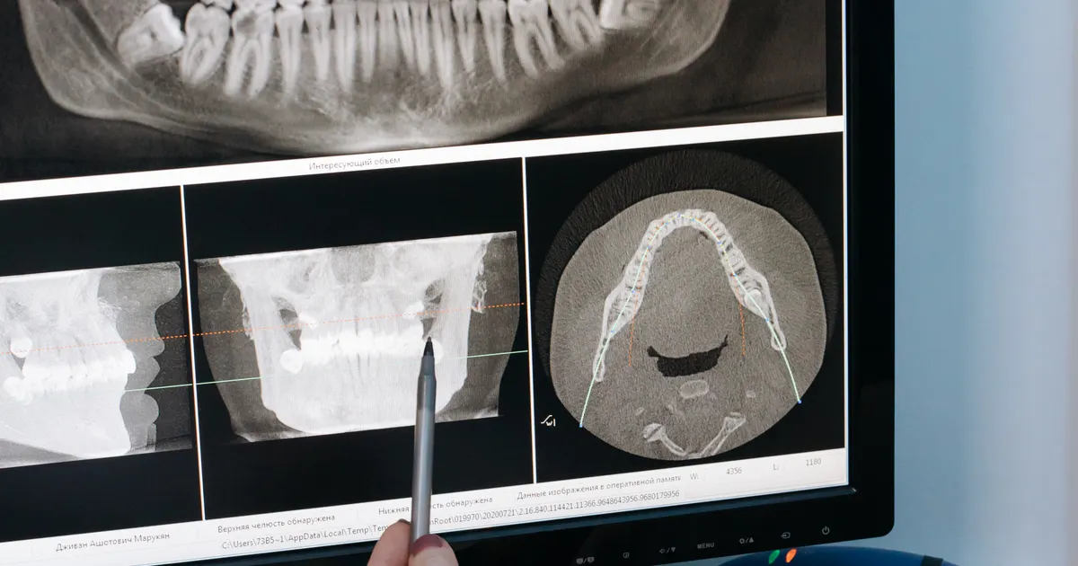
On an X-ray, a cyst usually appears as a round-to-oval dark (radiolucent) area at the tip of a tooth root or within the jawbone; its border is often clear and smooth, and there may be a thin white sclerotic line around it. However, not every dark area on an X-ray is a cyst — a granuloma, an abscess, a healing area and anatomical spaces can all produce a similar picture. When needed, the size, the border and the relationship to neighbouring structures are clarified with CBCT; the final diagnosis is made through clinical examination together with the histology result.
What does the research say? A radiolucent (light-transmitting) area on an X-ray may be a cyst, a granuloma, an abscess, a healing area or an anatomical space; according to a 2013 J Endod study, even with CBCT it is possible to distinguish a cyst from a granuloma only with limited accuracy, and a 2020 study shows that grey values serve as a supporting criterion. The final diagnosis is made through histological examination; according to the AAE/AAOMR position statement, CBCT is used, where indicated, to assess the size, the border and the relationship to neighbouring structures. Related: 3D diagnostics (CBCT) service · Can an infection in the jaw be seen on an X-ray? · Which conditions can be seen on CBCT?.

What is a cyst and why is it visible on an X-ray?
A dental cyst is a sac-like structure filled with fluid, with a wall made up of epithelial cells. As the cyst grows, it begins to dissolve the surrounding bone tissue — in scientific terms, a process called resorption. It is precisely this bone loss that allows X-rays to pass through that area more easily. As a result, that spot appears darker than the surrounding tissues on the X-ray film or screen.
To detect dental cysts in good time, a panoramic X-ray is the most widely used initial diagnostic method. In doubtful cases, CBCT (cone-beam computed tomography) is used for more precise information.
What does a periapical cyst look like on an X-ray?
The periapical cyst is the most frequently encountered type of cyst in X-ray diagnostics. This cyst forms at the tip of the tooth root — in the periapical region. That is where its name comes from: peri means around, apex means the root tip.
Its features on the X-ray image:
- A dark, round or oval shadow directly adjacent to the tip of the tooth root
- A smooth, clearly defined border — this indicates that the cyst is growing slowly
- A thin, whitish sclerotic rim around it — the bone's protective reaction
- Usually larger than 5 mm in size; but it can sometimes reach 2–3 cm
Please note: In the early stage, small periapical cysts look very similar to a granuloma. Sometimes only histological examination is enough to tell them apart. That is why it is important for the X-ray image to be read by an experienced specialist.
What does a jaw cyst look like on a panoramic X-ray?
Jaw cysts form a broad group: odontogenic cysts (related to a tooth), non-odontogenic cysts (arising from epithelial remnants) and developmental cysts (for example, a dentigerous cyst — a cyst that forms around an unerupted tooth).
The general picture on a panoramic X-ray:
- A wide, smooth-edged radiolucent (dark) area
- Large cysts may extend beyond the boundaries of several teeth
- In a dentigerous cyst, the crown of the unerupted tooth lies within the dark area
- Rapidly enlarging cysts can thin the bone plate and cause deformation
A panoramic X-ray shows the overall picture of the cyst, but it does not provide depth or three-dimensional information. To fill this gap, a CBCT examination is invaluable.
What does a dental cyst show on a CBCT image?
A CBCT examination of a dental cyst provides far more detailed information than a standard X-ray or a panoramic image. CBCT — cone-beam computed tomography — images the jaw in three planes (front-to-back, side, top-to-bottom).
The advantages of CBCT:
- Precise measurement: The width, height and depth of the cyst are measured with millimetre accuracy.
- Assessment of the bone wall: The proximity of the cyst to the nasal cavity, the lower jaw canal or the sinus is clearly visible.
- Effect on neighbouring teeth: It is determined which tooth roots extend into the area inside the cyst.
- Surgical planning: Based on the CBCT image, the surgeon draws up a precise plan before the operation.
In Baku, patients with a suspected cyst are in most cases advised to have a combination of both a panoramic X-ray and CBCT. Using these two examinations together both refines the diagnosis and optimises the treatment plan.
Not every dark area on an X-ray is a cyst — please note
It is not correct to see a dark area on an X-ray image and immediately say cyst. A number of other conditions can produce a similar picture:
| Condition | X-ray appearance | Distinguishing feature |
|---|---|---|
| Periapical granuloma | Dark, small round area | Usually smaller than 5 mm, less clearly defined edge |
| Dental abscess (acute) | Diffuse dark area in the periapical region | Indistinct edges, acute clinical symptoms |
| Normal anatomical structure | Mental foramen, sinuses | Constant location, symmetrical appearance |
| Ameloblastoma | Multilocular (soap bubble) appearance | Large, aggressive growth pattern |
| Ossifying fibroma | Mixed dark-and-white appearance | Areas of calcification within it |
For this reason, only a trained dental radiology specialist can read an X-ray image correctly. Self-assessment can lead to a wrong diagnosis — and to both unnecessary intervention and delayed treatment.
The importance of early diagnosis
Cysts often grow without pain. Even if the patient feels nothing, an X-ray examination taken every six months makes it possible to detect a cyst at an early stage on the image. What does early detection mean?
- Less bone loss
- A simpler surgical procedure
- Less risk for the neighbouring teeth
- A faster healing process
Large cysts, on the other hand, sometimes require the removal of several teeth, extensive bone resection and even bone grafting. A preventive X-ray examination is the most effective way to avoid these serious outcomes.
Frequently asked questions
Q: Is a cyst always visible on an X-ray? A: Very small cysts (2–3 mm) may not be visible on a standard X-ray. In such cases, CBCT is a more sensitive diagnostic option. A comprehensive assessment carried out together with a clinical examination gives the most reliable result.
Q: What does a periapical cyst look like on an X-ray? A: On an X-ray image, a periapical cyst appears as a round, dark (radiolucent) area adjacent to the tip of the tooth root. A smooth, sometimes whitish line (a sclerotic rim) can be seen around it.
Q: How does CBCT differ from a standard X-ray? A: CBCT gives a three-dimensional image. While a standard X-ray shows only a flat projection, CBCT clearly reveals the exact size of the cyst, its relationship with the bone and its effect on neighbouring structures.
Q: Does every dark area mean a cyst? A: No. A dark area on an X-ray may also be an abscess, a granuloma, a bone defect or a normal anatomical structure. A specialist assessment is essential for an accurate diagnosis.
Q: Can a cyst go away on its own? A: No. Dental cysts do not disappear spontaneously; on the contrary, they grow over time. Early intervention simplifies treatment and reduces bone loss.
Author: Dr. Bakhtiyar Aliyev, Dental Radiology Specialist | Smile by Dr. Bakhtiyar, Baku. This article has been reviewed by a dentist.
For a professional dental X-ray in Baku, get in touch with us — book online or give us a call.
Sources
- Evaluation of the reliability and accuracy of using cone-beam computed tomography for diagnosing periapical cysts from granulomas — Journal of Endodontics, 2013
- Accuracy of mean grey density values obtained with small field of view cone beam computed tomography in differentiation between periapical cystic and granulomatous lesions — International Endodontic Journal, 2020
- AAE and AAOMR Joint Position Statement: Use of Cone Beam Computed Tomography in Endodontics — 2015 Update — Oral Surgery, Oral Medicine, Oral Pathology and Oral Radiology, 2015 (AAE/AAOMR)
This article is for general information only and does not replace an individual diagnosis, examination or treatment. Consult a doctor for a decision about your case.