

Does a jaw infection show up on an X-ray?
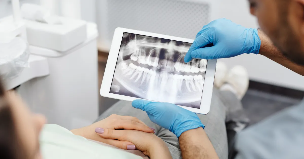
Yes — when an infection at the root tip dissolves bone, it shows up on an X-ray as a dark (radiolucent) area at the tip of the tooth root; however, at a very early stage, while mineral loss in the bone is still minimal, an ordinary 2D X-ray may not show any change. A periapical X-ray is the first choice (dose 2–5 µSv); according to the joint position statement of the AAE and the AAOMR, CBCT is added with a small FOV only in cases where 2D does not answer the question — an unclear diagnosis, complex anatomy, retreatment, surgical planning. The diagnosis is always made together with a clinical examination (biting, percussion, vitality tests); an X-ray on its own does not say «there is or is not an infection». This article is about inflammatory (periapical) findings; whether the dark area at the root tip is a cyst, and how cysts are told apart, is covered in a separate article: How does a cyst look on an X-ray?.
How does an X-ray detect infection?
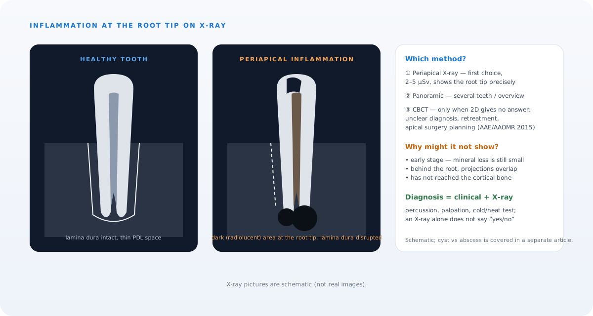
During a dental infection, that is an abscess, bacteria pass through the root canal to the tip of the root and from there into the surrounding bone tissue. When bone dissolves, that area absorbs less of the X-ray beam and appears on the image as a dark, transparent area — in medical terms a radiolucent zone. Healthy bone, on the other hand, absorbs more of the beam and appears light grey.
It is precisely this difference in contrast that allows the dentist to assess the presence, location and approximate size of the infection. That is why, when you feel toothache, swelling or heat, the first step is a clinical examination and, where indicated, a periapical X-ray.
What does a periapical abscess look like on an X-ray?
A periapical abscess is a collection of pus that gathers at the tip of a tooth root. On an X-ray, a dental abscess reveals itself through several characteristic signs:
- Periapical radiolucent area: a round or oval dark zone at the tip of the tooth root. This indicates the area where bone has dissolved.
- Widening of the periodontal space: the gap between the tooth root and the bone appears wider than normal.
- Disruption of the lamina dura: interruption or loss of the thin line of bone (the lamina dura) surrounding the root.
- Uneven appearance of the root canal: infection may have escaped from an untreated part of the canal.
In chronic (long-standing) abscesses these signs are clearer on the X-ray, because bone erosion has already developed. In acute (suddenly starting) abscesses, however, the process may not yet have reached the bone and the X-ray image can look “normal” at first glance. This is why a clinical examination must always complement the X-ray.
Which type of X-ray shows infection best?
Periapical X-ray (periapical radiograph)
Covering one specific tooth and the area around it, this image is the most widely used method for showing the early signs of infection. The dentist places this small film next to the tooth so that it fully covers the root tip of the suspected tooth.
Panoramic X-ray
A panoramic X-ray shows the whole jaw, all the teeth, the sinuses and the jaw joints in a single image. It is convenient for a broad overview, but it falls behind the periapical X-ray in fine detail. When infection is suspected in several teeth at once, a panoramic image may be the first choice.
CBCT — three-dimensional examination
CBCT (cone-beam computed tomography) images the jaw in three dimensions and shows periapical changes earlier and more precisely than 2D — but its dose is many times higher and, according to the AAE/AAOMR position statement, it is not routine; it is taken with a small FOV only for a question that 2D has not answered. This technology:
- Determines the exact location and extent of the infection,
- Shows how close the abscess is to the sinus cavity, the nerve canal or the neighbouring teeth,
- Reveals early-stage infections that are not visible on a standard X-ray,
- Creates a precise anatomical map for surgical planning.
At our clinic, CBCT examination is mainly taken where indicated before complex and repeat root canal treatment, apical surgery planning, implant planning and jaw surgery; for treating the infection, see: root canal treatment service.
Infections that do not show on an X-ray — what should be done?
Sometimes a patient feels severe pain, yet no change is visible on the X-ray. How is this possible?
There are several explanations:
- Early-stage infection: the process has not yet reached the bone and is confined to the soft tissue.
- The projection limitation of X-rays: a two-dimensional image can sometimes “mask” a pathology lying behind the root.
- Vertical bone loss: sometimes bone loss occurs vertically rather than horizontally; in this case it is difficult to see on a periapical X-ray.
- Odontogenic sinusitis: if the infection has spread into the upper jaw sinus, an ordinary dental X-ray is not enough.
In such cases, clinical tests first (percussion, cold/heat vitality testing, palpation) and, if needed, a second periapical image from a different angle, and — if the question remains — a small-FOV CBCT together give a more reliable result. For pain during and after root canal treatment, see: is root canal treatment painful.
What happens if infection is not addressed in time
If the diagnosis of a jaw infection is delayed, serious complications can develop:
- The infection can spread to the throat, the neck or the space beneath the eye (cellulitis, spread of the abscess).
- Bone tissue dissolves over a wider area (risk of osteomyelitis).
- Neighbouring teeth and dental implants can be damaged.
- There is a risk of systemic (whole-body) infection.
That is why, when toothache, facial swelling, heat or a feeling of pressure in the jaw appears, it is important to seek help as soon as possible. In Baku, this examination can be carried out in professional conditions.
Frequently asked questions
Q: Does a jaw infection always show on an X-ray? A: In most cases — yes. However, at a very early stage, while mineral loss in the bone is still minimal, a 2D X-ray may not show any change. In such cases clinical tests are key; CBCT is added only if the question remains.
Q: How does a dental abscess look on an X-ray? A: It appears as a dark, round or oval area at the tip of the tooth root — this is called a periapical radiolucent zone. In chronic abscesses this area is larger and clearer.
Q: Can the difference between a periapical abscess and a cyst be seen on an X-ray? A: On a standard X-ray the two look very similar. A CBCT examination, and sometimes a biopsy, is needed to tell them apart precisely.
Q: How does CBCT differ from a standard X-ray? A: CBCT images the jaw in three dimensions and shows the exact location and size of the infection and how close it is to the surrounding nerves and sinuses. A standard X-ray, on the other hand, gives a two-dimensional, that is a “flat”, image.
Q: Where should I go in Baku for an X-ray? A: Standard periapical X-rays, panoramic X-rays and CBCT examination are all available at the Smile by Dr. Bakhtiyar clinic. Online booking can be done conveniently through our website.
Author: Dr. Bakhtiyar Aliyev, Dental Radiology Specialist | Smile by Dr. Bakhtiyar, Baku. This article has been reviewed by a dentist.
For professional dental X-rays in Baku, get in touch with us — book online or give us a call.
Sources
- AAE and AAOMR Joint Position Statement: Use of Cone Beam Computed Tomography in Endodontics 2015 Update — Oral Surgery, Oral Medicine, Oral Pathology and Oral Radiology, 2015
- Outcome measures to assess the effectiveness of endodontic treatment for pulpitis and apical periodontitis for use in the development of European Society of Endodontology S3-level clinical practice guidelines — International Endodontic Journal, 2021 (ESE)
- Patient risk related to common dental radiographic examinations: the impact of 2007 International Commission on Radiological Protection recommendations regarding dose calculation — Journal of the American Dental Association, 2008
This article is for general information only and does not replace an individual diagnosis, examination or treatment. Consult a doctor for a decision about your case.