

Why is dental photography important in treatment planning?
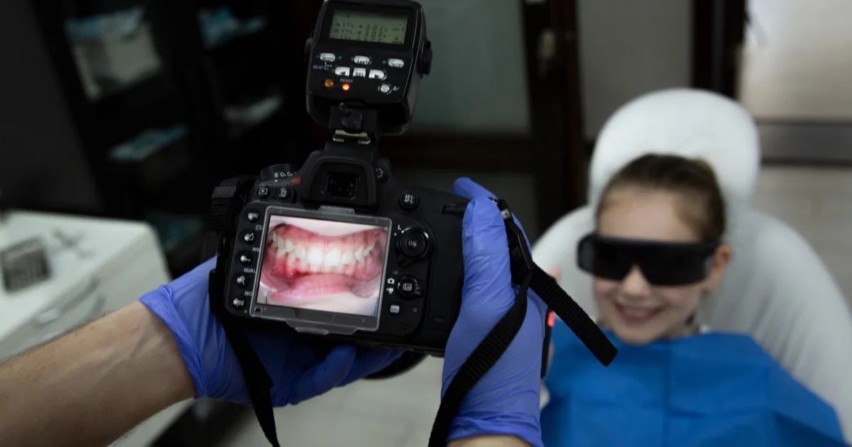
Dental photography is the documentation of the teeth, gums and smile using a standardised photographic protocol. These photographs serve four main purposes: an objective record of the starting situation, smile design and treatment planning, communicating shade and shape information to the laboratory, and comparative monitoring of treatment progress. In other words, the aim is not a nice picture — it is precise clinical information.
What is dental photography?
It is a clinical photography protocol carried out according to defined rules: the same angles, the same series of distances, intraoral images taken with retractors and mirrors, facial and smile frames, and photographs taken with a shade guide. Standardisation matters because photographs taken at different times need to be properly comparable with one another. The professional literature, for example in cases of trauma, describes clinical photography as an integral part of documentation.
How it is used: four main roles
1. Diagnostic documentation
The situation before treatment — existing restorations, wear, gum levels, shade characteristics — is recorded in a photographic series. This is both part of the clinical record and, years later, the precise answer to the question how did it look before?
2. Planning and smile design
Facial and smile photographs are the raw material of digital smile design: facial reference lines, the lip line and tooth display are analysed precisely from the photographs, and the design is then combined with the intraoral scan. Systematic reviews highlight the role of the photo-based digital design process in clarifying the patient's expectations and in shared decision-making.
3. Communication with the laboratory: shade and shape
Technicians do not see the patient — they get to know the tooth from the photograph. A photograph taken with a shade guide conveys to the dental technician the depth of the shade, the zones of translucency and the surface texture. Studies show that using photographic and instrumental methods alongside the visual method increases reliability in shade selection; we have written about this topic separately: how is tooth shade selected?
4. Comparison and monitoring
Photographs of the interim stages show the progress of treatment objectively: the mock-up trial, the temporary restorations, the final result. The before-and-after comparison is based not on impressions but on frames taken under identical conditions.
In which treatments is it particularly important?
- Veneers and aesthetic restorations — every decision about shade, shape and proportion goes through photographic analysis.
- Prosthodontic treatment and large restorative cases — an exchange of photographs with the laboratory at every stage.
- Gum aesthetics and long-term monitoring — comparison of changes in level.
- Trauma and unusual clinical cases — medico-legal documentation of the situation.
What does the patient gain?
- A transparent decision — you can see what will be changed and why, on your own photographs.
- More precise laboratory work — the technician receives standardised photographic information rather than words.
- Objective comparison — the result is assessed as documented, not as remembered.
- An archive — even years later, the starting situation can be reviewed.
Is a phone camera not enough?
It is an interesting question and one that is studied in the literature. Comparative studies show that modern smartphone cameras can be used for some shade assessment tasks under controlled conditions, but the result depends heavily on lighting and standardisation. The value of a clinical protocol lies not in the brand of the camera but in reproducible conditions: the same light, the same angle, a shade guide. That is why photography in the clinic is not casual picture-taking but a matter of protocol.
How is the photographic protocol set up at Smile by Dr. Bakhtiyar?
At our clinic in Baku, photographic documentation is a standard part of planning in aesthetic and prosthodontic work: before treatment, a facial, smile and intraoral series is taken; at the design stage these photographs are combined with digital smile design and the TRIOS scan; and frames with a shade guide are sent to the laboratory. Dr. Bakhtiyar Aliyev's list of training includes a specialist course in dental photography; he makes the design decisions personally. Together with our other technologies: Technology page.
Frequently asked questions
Where do the photographs go? Is confidentiality protected? The photographs are part of the clinical record and are kept in the patient's file. Public sharing is only possible with the patient's separate consent.
How long does the photography take? A standard series usually takes 10–15 minutes. No separate preparation is required.
Are the retractors uncomfortable? There may be a slight feeling of tension, but they do not cause pain; each frame takes a few seconds.
Does this replace an X-ray? No. Photographs only record the visible surfaces; for the inside of the tooth and for bone, an X-ray or CBCT is needed — these are tools with different purposes.
Is it taken for every patient? The extent depends on the type of work: a full series for aesthetic and prosthodontic cases, and only the necessary frames for smaller treatments.
Clinical note: This article is for general information and does not replace individual advice from your dentist. To discuss aesthetic planning, book an appointment.
Sources
- Clinical photography and documentation after traumatic dental injuries — Dental Traumatology, 2024
- Comparative Evaluation of Shade Selection Reliability: Visual Assessment, Intraoral Scanning, and Photographic Techniques — International Journal of Dentistry, 2026
- Can smartphone cameras be a viable alternative to digital single-lens reflex (DSLR) cameras for dental shade assessment? — Evidence-Based Dentistry, 2026
- Digital Smile Design and Patient-Centered Outcomes in Esthetic Restorative Dentistry: A Systematic Review — Cureus, 2025
This article is for general information only and does not replace an individual diagnosis, examination or treatment. Consult a doctor for a decision about your case.