

Gum aesthetics around implants: thin and thick gums, papilla and grafting
The appearance of an implant is often decided not by the implant itself, but by the gum around it. If the gum is thin, the metal underneath casts a grey shadow and the margin recedes over time; thick, keratinised (firm, chewing-type) gum both hides the colour and stays more stable in place. That is why, in the front region, the plan often includes thickening the gum (a connective tissue graft) and shaping it with a temporary crown. The result is individual and requires care over the years.
Key points
- The gum biotype (whether it is thin or thick) is one of the main factors for colour, margin stability and papilla around an implant.
- The evidence on how keratinised gum affects long-term health is uncertain, but its advantage in terms of ease of cleaning and patient comfort is noted (EFP/AAP consensus, 2018).
- A connective tissue graft can thicken the gum; how long the result lasts depends on individual factors.
- The papilla (the triangle of gum between the teeth) is determined by the level of the neighbouring tooth and of the bone between implants — surgically creating it is limited.
- Shaping the gum with a temporary crown is the main tool for building a natural emergence profile around an implant.
We are not repeating here what an implant is and how it integrates — there is the what is a dental implant article for that. The general plan for implant placement in the front region (bone, position, prosthesis) is explained separately in the implant placement in the aesthetic zone piece. Here we talk only about the soft tissue — the gum.
How does the gum around an implant differ from the gum around a natural tooth?
Around a natural tooth, the gum is attached to the root surface by fibres. Around an implant there is no such attachment: the gum only adheres to the implant and the abutment (the intermediate part connecting the implant to the crown), with the fibres running parallel to the surface. According to the EFP/AAP consensus report, healthy peri-implant gum is on average 3–4 mm in height and may be covered either by keratinised (firm, chewing-type) or by non-keratinised (soft, mobile) epithelium.
The practical consequence is this: the gum around an implant is more fragile than around a natural tooth. The blood supply is weaker, and it is more sensitive to mechanical trauma and bacterial plaque. That is why, in the front region, the gum plan is not left to chance.
Thin and thick gums: what is a biotype?
Biotype (now more often called phenotype) refers to the thickness of the gum and the width of the keratinised band. Put simply:
- Thin phenotype: the gum is translucent, the probe shows through it, the teeth are long and conical, the papillae narrow. This kind of gum is prone to recession and lets the grey colour of the underlying abutment show through.
- Thick phenotype: the gum is dense and opaque, the teeth close to square, the papillae full. This kind of gum is more resistant to trauma and hides colour better.
This difference affects not only appearance but also the bone. In a 1-year prospective controlled study by Linkevicius and colleagues (19 patients, 46 implants), in sites with thin gum (≤2 mm) bone loss at the implant neck averaged 1.61 mm on the mesial side, compared with 0.26 mm in sites with thick gum; on the distal side the figures were 1.28 mm and 0.09 mm respectively. The authors regard initial gum thickness as an important factor affecting the stability of the bone around the implant. We cover the topic of bone level separately in the bone loss around implants article.
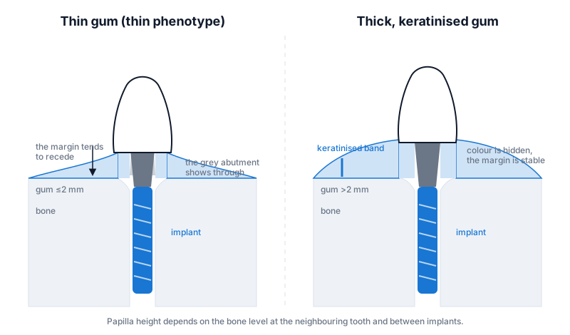 On the left, thin gum: the colour of the abutment shows through and the margin is prone to recession. On the right, thick, keratinised gum: the colour is hidden and the margin is stable.
Why is keratinised gum needed?
Keratinised gum is the firm, pale pink, immobile band around the tooth. Below it begins the mobile, soft mucosa. When this band is narrow or absent around an implant, the gum hurts when the brush and floss touch it, the patient avoids cleaning that area, and plaque builds up.
The evidence is cautious on this point. The EFP/AAP consensus report calls the effect of keratinised gum on long-term peri-implant health uncertain, but notes that it may be advantageous in terms of patient comfort and ease of plaque removal. The same document lists thin soft tissue, absence of keratinised tissue, incorrect implant position, absence of labial (lip-side) bone and surgical trauma among the main causes of gum recession.
In the 2018 systematic review and meta-analysis by Thoma and colleagues, implant sites that received a soft tissue graft were compared with sites without a graft: in the grafted group bleeding scores were lower and the bone level at the implant neck was somewhat more stable (a mean difference of about 0.1 mm over the observation period of the studies). The difference is small, but the direction favours grafting; for autogenous grafts (taken from the patient's own palate) the results were clearer.
Papilla: why is the triangle between the teeth lost, and does it come back?
The small triangle of gum between the front teeth — the papilla — is important for a natural-looking smile. When it is lost, a black triangle remains in its place. And this is the question patients ask most often: can the papilla be restored?
An honest answer is needed here. According to the consensus report, the height of the papilla between an implant and a natural tooth is determined by the level of the periodontal (tooth-supporting) tissues of the neighbouring tooth, while the papilla between two implants is determined by the crest of bone between the implants. In other words, the papilla is suspended from the support beneath it. If that support is absent, building a full papilla with soft tissue surgery alone is difficult.
That is why the papilla plan is set out from the start:
- Preserving the bone of the neighbouring tooth during extraction (atraumatic extraction, socket preservation).
- Placing the implant at a sufficient distance from the neighbouring tooth and from a second implant — the positioning rules are given in the 3D implant position piece.
- Shaping the contact point of the crown so that the space for the papilla is neither too open nor too compressed.
Gum thickening: what is a connective tissue graft?
A connective tissue graft (CTG) is a thin piece of tissue taken from the inner layer of the palate and placed around the implant, under the gum. The aim is to thicken the gum, mask the colour of the abutment and fill out the labial contour. The palatal site heals in 1–2 weeks; as an alternative, collagen matrices (tissue substitutes) are also used, but the evidence for gains in thickness favours autogenous grafts.
According to the 2014 systematic review by Thoma and colleagues, a graft (free gingival graft, connective tissue graft or collagen matrix) combined with an apically positioned flap/vestibuloplasty to increase the keratinised band increased keratinised tissue by 1.4–3.3 mm over the follow-up period of the studies; for soft tissue thickness, autogenous grafts gave better results than ungrafted controls. The clinical guidance is simple: if the keratinised band needs to be increased, a free gingival graft; if thickness and colour are needed in the front region, a connective tissue graft.
When is the graft done? There are three options:
- On the same day as the implant — if the gum is thin, so that surgery is completed in one go.
- At implant exposure (second stage) — the gum has already healed and the contour is easy to assess.
- After the crown has been fitted — when recession or a grey shadow appears. This is technically the most difficult option, because the tissue has already formed.
In the front region the first or second option is usually chosen; which one it will be is decided on the basis of the bone contour on 3D CBCT and the clinical thickness of the gum.
Shaping the gum with a temporary crown
Gum is a tissue with a memory: whatever shape you give it, it heals in that shape. What is placed on the implant first is not the permanent crown but a temporary one (usually PMMA or composite). Its neck portion is altered gradually beneath the gum: material is added on one side so that the gum is pushed away, and reduced on the other so that the tissue moves down. This process takes from a few weeks to a few months, and the result is a transition from the round neck of the implant to the triangular-oval neck shape of a natural tooth — the emergence profile.
When the permanent crown is made, this shape is transferred precisely to the laboratory; in the clinic a customised impression or an intraoral scanner is used for this. A custom abutment (not a standard one, but made to match the shape of the gum) is helpful at this stage. The materials themselves — zirconia, titanium, ceramics — are the subject of the screw-retained or cement-retained prosthesis and materials articles.
How stable is the result in the long term?
Patients rightly ask: the graft was done, but will it not recede again in a few years? The honest answer: soft tissue around an implant changes somewhat over the years, and the extent of this change is individual. The factors affecting stability are:
- Bone support. If the labial bone is thin or absent, the gum may not stay in place on its own for long; although the consensus report calls the evidence on the long-term role of the buccal bone uncertain, this is taken into account in planning.
- Implant position. Around an implant placed too far towards the lip or too shallow, the gum recedes sooner.
- Hygiene and inflammation. Inflammation around the gum (peri-implant mucositis) is the beginning of tissue loss; the topic is covered in the what is peri-implantitis piece.
- Brushing technique. A hard brush and horizontal movements mechanically pull thin gum downwards.
- Smoking. It impairs the blood supply of the soft tissue and makes it harder for the graft to take.
That is why the annual check-up after an implant is not only an X-ray, but also a record of the level of the gum margin and of bleeding. This routine is described in the long-term care after implants article. When dental implantation is planned, assessment of the soft tissue in the front region is part of the first consultation.
Frequently asked questions
The gum looks grey after the implant was placed. Can this be corrected? The cause is usually the titanium abutment showing through thin gum. A solution is possible in three ways: thickening the gum with a connective tissue graft, replacing the abutment with a zirconia or tooth-coloured coated version, or combining the two. Which way is suitable is determined by the gum thickness and the position of the implant.
Is taking tissue from the palate painful? The donor site on the palate is usually more bothersome than the implant site and stays sensitive for 1–2 weeks. A protective plate or a collagen dressing makes this period easier. Tissue substitutes remove the need for a palatal site altogether, but the gain in thickness is usually less than with an autogenous graft.
Will the black triangle between the implant and the neighbouring tooth fill in? The papilla depends on the bone level of the neighbouring tooth. If the bone at the neighbouring tooth is intact, the papilla fills in considerably with a correct contact point and shaping with a temporary crown. If bone has been lost, complete filling is not expected; in such cases the gap is masked by the shape of the crown.
If there is little keratinised gum, will the implant definitely fail? No. The evidence does not directly link the absence of a keratinised band with implant loss. But cleaning becomes more difficult and the gum becomes sensitive; so if mobile mucosa extends right up to the implant, grafting is discussed.
Can the temporary crown stage be skipped? On the back teeth, sometimes yes. In the front region, however, the temporary crown is the main tool that shapes the gum; skipping it can lead to an uneven appearance of the gum margin around the permanent crown.
Conclusion
Aesthetics around an implant are determined by the thickness of the soft tissue, the presence of a keratinised band and the bone support that holds the papilla. Thin gum creates problems both with colour and with stability; a connective tissue graft and shaping with a temporary crown are the two main means of reducing these risks. No method locks in the result for life — annual check-ups and correct hygiene are part of the plan. To assess an individual situation, the thickness of the gum, the width of the keratinised band and the bone contour on 3D imaging are reviewed together at the consultation.
Sources
- Peri-implant diseases and conditions: Consensus report of workgroup 4 of the 2017 World Workshop on the Classification of Periodontal and Peri-Implant Diseases and Conditions — Journal of Clinical Periodontology, 2018 (EFP)
- Efficacy of soft tissue augmentation around dental implants and in partially edentulous areas: a systematic review — Journal of Clinical Periodontology, 2014
- Effects of soft tissue augmentation procedures on peri-implant health or disease: A systematic review and meta-analysis — Clinical Oral Implants Research, 2018
- The influence of soft tissue thickness on crestal bone changes around implants: a 1-year prospective controlled clinical trial — International Journal of Oral and Maxillofacial Implants, 2009
This article is for general information only and does not replace an individual diagnosis, examination or treatment. Consult a doctor for a decision about your case.