

Bone loss around an implant: normal remodelling or a problem?
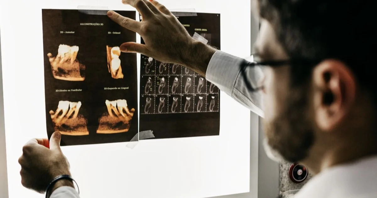
A certain amount of bone change at the implant neck during the first year is expected — this is called physiological remodelling, and the EFP/AAP consensus does not consider it a disease. What is concerning is progressive loss that goes beyond this initial change and continues over the years, usually accompanied by bleeding gums and increasing probing depth. The only reliable way to tell the two apart is to compare the baseline X-ray taken when the prosthesis is fitted with later follow-up X-rays.
Key points
- A slight drop in bone level during the first year does not mean the implant is dissolving; the extent varies with implant design, gum thickness and the surgical protocol.
- According to the consensus, if no earlier X-ray is available, a bone level 3 mm or more below the top edge of the intraosseous part of the implant plus bleeding on probing points towards peri-implantitis.
- In meta-analyses, platform switching (an abutment narrower than the implant) preserved on average 0.4–0.6 mm more bone at the neck.
- Thin gums (2 mm or less) are associated with greater bone loss in the first year.
- There is usually no pain; this is why follow-up X-rays matter even in the absence of symptoms.
We will not repeat here how an implant fuses with bone — the article what is a dental implant explains that. The question here is narrower: when a follow-up X-ray shows that the bone has dropped a little, what does that mean and when should you be concerned?
Physiological remodelling: what happens in the first year?
After the implant is placed and loaded with a prosthesis, the bone at the neck of the implant adapts to its new situation. For the gum to attach to the implant, the body builds a soft-tissue cuff of a certain height; if there is no room for this cuff, the bone recedes to make space. This is called physiological remodelling.
The EFP/AAP 2017 World Workshop consensus report (Berglundh et al., 2018) states this plainly: the degree of physiological remodelling after implant placement is variable, and it is precisely this remodelling that determines the expected bone level around a healthy implant; its extent depends on a number of local and systemic factors. The same report recommends two X-rays to the dentist: one when the prosthesis is completed (baseline) and another after the loading period — in order to record the level after remodelling has finished as the reference point. All subsequent comparisons are made against this second X-ray.
The consensus also adds a caution: extensive bone loss occurring during the remodelling period may also be a sign of peri-implantitis that began in that same period. In other words, the phrase it is the first year, this is normal only holds when the loss is limited and there are no signs of inflammation.
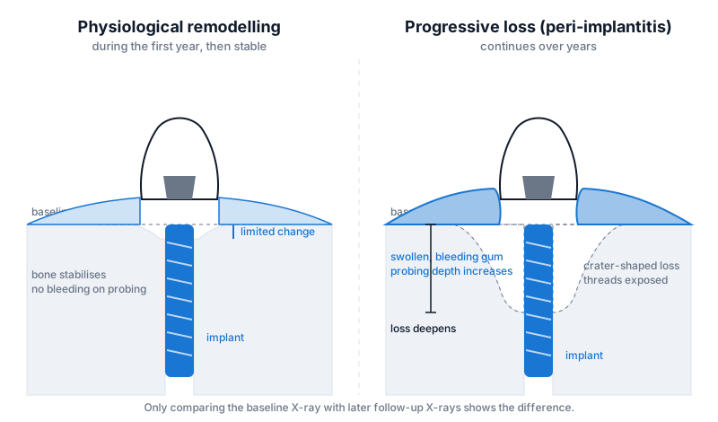 Left: limited bone change that stabilises after the first year. Right: saucer-shaped (crater) loss continuing over the years, with inflamed gums.
Pathological loss: when is it called a problem?
The clinical name for progressive loss is peri-implantitis. The definition proposed by the consensus report for everyday practice requires three conditions together: bleeding and/or pus on gentle probing; increased probing depth compared with the previous examination; and bone loss beyond the initial remodelling.
If no earlier X-ray is available (for example, the implant was placed elsewhere and the records are not accessible), the consensus offers another criterion: bleeding/pus on probing plus a probing depth of 6 mm or more plus a bone level 3 mm or more below the most coronal point of the intraosseous part of the implant. For epidemiological studies, it is recommended that measurement error (on average 0.5 mm) be taken into account — meaning that differences of 0.3–0.5 mm mean nothing on their own.
The key table distinguishing these two situations:
| Criterion | Physiological remodelling | Progressive (pathological) loss |
|---|---|---|
| Timing | Mainly the first year, then stabilises | Continues over the years |
| Shape on X-ray | Small, flat or slightly saucer-shaped | Deepening crater, sometimes one-sided |
| Bleeding on probing | Absent | Present, sometimes with pus |
| Probing depth | Stable | Increasing |
| Gums | Quiet | Red, swollen or receded |
| What to do | Keep the reference X-ray, monitor | Find the cause, start treatment |
The treatment of peri-implantitis itself is not the subject of this article; it is explained separately in what is peri-implantitis. For implant mobility, pain and other serious signs, there is the article signs of implant failure.
Factors affecting bone loss
Platform switching and connection design
Platform switching means the abutment is slightly narrower than the top platform of the implant. As a result, the implant-abutment junction (the microgap and the bacteria that collect there) is moved inwards, away from the bone margin. Two meta-analyses have put figures on this: in the 2010 review by Atieh et al. (10 studies, 1239 implants), marginal bone loss around platform-switched implants was on average 0.37 mm less than around platform-matched implants, with no difference found in terms of implant loss. In the 2012 review by Annibali et al. (10 randomised studies, 435 patients, 993 implants), the difference at patient level was 0.55 mm; again no difference was found in implant success rates. In other words, platform switching does not rescue an implant, but it does preserve the bone at the neck somewhat better. The connection types of the systems used in the clinic are compared in the article implant systems.
Gum thickness
If the gum is thin, room for the soft-tissue cuff mentioned above is made at the expense of the bone. In the 1-year prospective controlled study by Linkevicius et al. (19 patients, 46 implants), up to 1.45 mm of bone loss was recorded at the implant neck in the first year at sites with gums 2 mm thick or less; at sites with thick gums, the loss was less than 0.3 mm. This is one reason why thickening the gum in the front region is discussed — the topic is covered in gum aesthetics around an implant.
Implant position and depth
In an implant placed too shallow, the rough surface emerges below the gum; in one placed too deep, remodelling eats away more bone. Placement too close to a neighbouring tooth or to a second implant loses the crest of bone in between. These rules are set out in the article 3D implant position.
The prosthesis and residual cement
Cement left below the gum creates a surface for bacteria; the consensus report lists this among the factors with limited evidence linked to peri-implantitis. As for bone level, the 2016 meta-analysis by Lemos et al. (20 studies, 8989 implants, mean follow-up 65 months) produced an interesting result: marginal bone loss with cemented prostheses was on average 0.19 mm less than with screw-retained prostheses. In other words, the type of construction alone does not determine the bone; proper removal of the cement and a shape that allows for hygiene matter more. In detail: screw-retained or cemented prosthesis.
Load and bruxism
Excessive chewing load — especially in people with a clenching habit — increases stress at the implant neck. The consensus report regards whether occlusal loading is a risk indicator for peri-implantitis as not yet established; that is, the link is suspected but not proven. In practice, in a patient with bruxism the shape of the crown and a night guard become part of the plan.
Inflammation and general factors
Bacterial plaque is the main cause; smoking, uncontrolled diabetes and a history of periodontitis are explained as factors that accelerate bone loss in the article smoking, diabetes and implant outcomes.
How is X-ray monitoring carried out?
Bone level is measured with an intraoral periapical X-ray; panoramic and 3D CBCT images are less accurate for this purpose (3D is added only when the shape of the crater needs to be seen). For proper monitoring:
- Baseline X-ray — on the day the prosthesis is fitted.
- Reference X-ray — about one year after loading; remodelling is considered complete.
- Follow-up X-ray — at intervals set by the dentist (individual, based on risk factors); taken each time at the same angle, with the parallel technique, so that millimetres can be compared.
Alongside the X-ray, probing depth and bleeding are recorded at every check-up, because the gums give warning before bone loss becomes visible on an X-ray. This routine is covered in detail in long-term care after an implant. When dental implantation is being planned, taking and keeping a baseline X-ray is a standard step at the clinic — later comparison is only possible with it.
When should you be concerned?
Contact your dentist ahead of schedule if:
- the gum around the implant bleeds, swells or discharges pus;
- the gum has receded and the threads of the implant are visible;
- discomfort has appeared in that area when brushing or with cold water (the implant itself has no sensation, but inflamed gums do);
- the bone level on a follow-up X-ray has clearly changed compared with the previous one;
- the crown moves or the screw loosens frequently.
The absence of pain is not reassuring: peri-implantitis runs a painless course for a long time. That is why the main message of this article is simple — keep your X-rays, come for your check-ups, and do not ignore bleeding.
Frequently asked questions
The first thread of the implant is visible on the X-ray. Does this mean the implant is lost? No, not on its own. With many implant designs, the bone may recede as far as the first thread during the first year and then remain stable. What matters is the trend: if the level stays the same on the next X-ray and the gums are quiet, this counts as remodelling.
Does lost bone come back? Bone lost through physiological remodelling is not restored, but it stays stable. With crater-type loss related to peri-implantitis, partial regrowth is possible in some cases with regenerative techniques once the inflammation has been stopped; this depends on the shape of the defect and is a separate treatment topic.
If I choose a platform-switched implant, will I have no bone loss? Meta-analyses show a difference of around half a millimetre on average; that is less loss, not zero loss. Gum thickness, position and hygiene are equally important.
How often should I have an X-ray? With a stable implant and low risk the interval is longer; with bleeding, smoking or a history of periodontitis it is shorter. The specific interval is set by the dentist based on the findings at check-ups; there is no standard universal figure.
Is a panoramic X-ray enough? For a general overview yes, for millimetre comparison no. An intraoral X-ray taken with the parallel technique is used to monitor bone level.
Conclusion
Limited bone change at the implant neck during the first year is remodelling and, according to the consensus, part of a healthy situation; progressive loss accompanied by bleeding and deepening probing depths, on the other hand, is peri-implantitis. The two can only be told apart with a baseline X-ray and regular check-ups. Platform switching, thick gums, correct positioning and a prosthesis that is either cement-free or properly cleaned all help to protect the bone, but none of them replaces monitoring. To have your individual situation assessed, it is helpful to bring your previous X-rays with you to your consultation.
Sources
- Peri-implant diseases and conditions: Consensus report of workgroup 4 of the 2017 World Workshop on the Classification of Periodontal and Peri-Implant Diseases and Conditions — Journal of Clinical Periodontology, 2018 (EFP)
- Platform switching for marginal bone preservation around dental implants: a systematic review and meta-analysis — Journal of Periodontology, 2010
- Peri-implant marginal bone level: a systematic review and meta-analysis of studies comparing platform switching versus conventionally restored implants — Journal of Clinical Periodontology, 2012
- The influence of soft tissue thickness on crestal bone changes around implants: a 1-year prospective controlled clinical trial — International Journal of Oral and Maxillofacial Implants, 2009
- Evaluation of cement-retained versus screw-retained implant-supported restorations for marginal bone loss: A systematic review and meta-analysis — Journal of Prosthetic Dentistry, 2016
This article is for general information only and does not replace an individual diagnosis, examination or treatment. Consult a doctor for a decision about your case.