

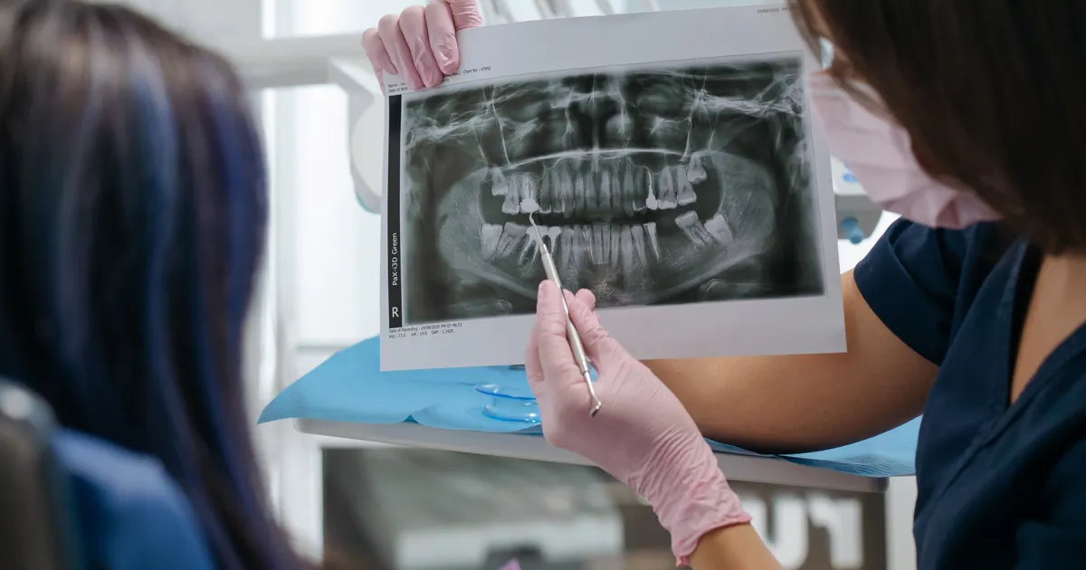
An occlusal X-ray is a wide intraoral X-ray taken by placing the sensor or film inside the mouth parallel to the occlusal (biting) surface of the upper or lower jaw. It is about four times larger than a standard periapical film and gives a broad view of several teeth, the alveolar ridge and the palatal bone in a single frame. It is most often used to see unerupted (impacted) teeth, extra (supernumerary) teeth, cysts and palatal pathology in children.
What does the research show? Among intraoral techniques, the occlusal X-ray covers the widest area, yet it is still a 2D method: it is useful for the initial assessment of the position of hidden teeth, cysts and bone changes in the midline of the jaw, salivary stones and fractures, but it does not show depth relationships (International Endodontic Journal, 2009). According to the AAOMR selection criteria, the imaging method is chosen according to the clinical question: a panoramic X-ray if a broad overview is needed, CBCT if a three-dimensional relationship is needed — the occlusal X-ray is not a replacement for them but an addition. The dose is low (a single intraoral exposure is in the microsievert range), but every image is justified by an indication. Related: 3D diagnostics (CBCT) service · What is a periapical X-ray? · What is a panoramic X-ray?.
Why is the occlusal X-ray a specific technique?
Unlike standard intraoral X-rays (periapical, bitewing), during an occlusal X-ray the sensor is pressed against the biting surface horizontally rather than upright between the teeth. The patient gently bites down on the sensor and the X-ray beam is angled downwards from above for the upper jaw, or upwards from below for the lower jaw. As a result, the image does not show just one or two teeth — it covers a wide region, including the buccolingual (cheek-to-tongue) direction of the bone.
The main advantage of this technique is that it provides a valuable image when placing a sensor becomes difficult in very young children, or when a wide-area lesion (a cyst, an extra tooth, unmineralised cementum and so on) is being looked for.

When is an occlusal X-ray taken?
The clinical indications are as follows:
- Locating unerupted (impacted) teeth: especially to determine the position of canines and central incisors within the palate.
- Extra (supernumerary) and mesiodens teeth: additional teeth hidden between the front incisors.
- Cysts and palatal pathology: enlarged dentigerous (follicular) cysts, radicular cysts, palatal tumours.
- Salivary and duct stones (sialoliths): where a stone is suspected in the duct of the submandibular (below the lower jaw) salivary gland.
- Trauma in children: to see root fractures in the front teeth and cracks in the alveolar bone.
- Cleft palate assessment: to follow the structure of the palate with a wide view.
- Displaced teeth and labial-palatal position: to determine whether a tooth is tilted towards the cheek or the palate.
How many types of occlusal X-ray are there?
Three main techniques are used in practice:
- Anterior occlusal (upper jaw): covers the front part of the upper jaw and the central incisors. It is the main choice for extra teeth and unerupted canines.
- Lateral occlusal (upper jaw): gives a wide cheek-to-palate view of the premolar-molar region.
- Anterior occlusal (lower jaw): shows the bone structure behind the lower incisors and the salivary gland duct.
- 90 degrees (mandibular true occlusal): a full view of the floor of the lower jaw, for assessing sialoliths and enlarged cysts.
How is an occlusal X-ray taken?
The procedure is very short:
- The sensor or film is placed in the mouth inside a special paper wrapping. The patient bites comfortably on the sensor — it causes no pain.
- The camera of the unit is angled at 65–70 degrees and directed from the forehead for an upper anterior occlusal image, and from under the chin for a lower occlusal image.
- An exposure of a few seconds — and the image comes through from the digital sensor to the computer.
- The result appears on the screen straight away.
A small-sized sensor is used for children; to avoid triggering the gag reflex, the sensor is placed quickly and in the correct position.
What does an occlusal X-ray show?
The image may show:
- The palatal structure of the upper jaw and buccolingual spread;
- Movement and positional change of the floor of the lower jaw;
- The exact location of unerupted and buried teeth;
- The borders and mineralisation of cysts and tumours;
- Sialoliths (salivary stones);
- Cracks in the alveolar bone and trauma;
- Foreign bodies in the palatal spaces.
However, an occlusal X-ray does not provide 3D depth information. For the exact buccal-palatal position of a tooth, bone thickness and complex anatomical relationships, CBCT (3D tomography) is superior.
The difference between an occlusal X-ray and other X-rays
- Occlusal vs periapical: a periapical image clearly shows the root of one or two teeth; an occlusal image shows a wide area. What is a periapical X-ray?
- Occlusal vs bitewing: a bitewing images only the crowns of the back teeth and the contact area; an occlusal image covers a whole jaw segment. What is a bitewing X-ray?
- Occlusal vs CBCT: an occlusal image is 2D and the dose is low; CBCT is 3D but requires a higher dose. In children and in cases of mild suspicion, the occlusal X-ray may be the first choice.
Occlusal X-rays in children — why are they so useful?
The occlusal X-ray has a special place in paediatric dentistry, because:
- In a young child the mouth is small, so it is difficult to position a periapical sensor correctly;
- Several teeth and bone structures are imaged in a single frame — this reduces the dose;
- It is more comfortable for children with a strong gag reflex;
- Problems with extra teeth and unerupted permanent teeth during the baby (primary) teeth period are easily detected.
More on the topic of dose: Are dental X-rays safe for children?.
Radiation dose
The dose of a digital occlusal X-ray is around 0.005–0.009 mSv — close to the natural radiation of a single day. A lead apron and a thyroid collar are used; in children the exposure is separately shortened according to a paediatric protocol.
Before and after the procedure
- No preparation is needed.
- Remove metal objects from the jaw area.
- You can return to normal life immediately after the procedure.
Conclusion
The occlusal X-ray is an intraoral tool that is limited like the periapical and bitewing, yet gives the big picture over a wide area. It is especially invaluable for unerupted teeth, extra teeth, cysts and palatal pathology in children. In digital format the dose is low and the result is instant.
In Baku, you can have occlusal, periapical and CBCT X-rays for both children and adult patients in our single digital X-ray room.
Book an appointment: Online appointment form
Frequently asked questions (FAQ)
1. Do I need to come on an empty stomach for an occlusal X-ray?
No, no special preparation is needed. The sensor is placed inside the mouth and the procedure takes a few seconds.
2. Is an occlusal X-ray harmful for a child?
The dose is extremely low — close to the natural background radiation of a single day. When carried out with protective shielding and on the basis of an indication, the risk in children is considered very low.
3. What is the difference between an occlusal X-ray and CBCT?
An occlusal X-ray is a 2D intraoral image showing a wide area. CBCT, on the other hand, is a three-dimensional scan that gives the exact volume of the bone. In complex situations, CBCT is recommended.
4. What should be done if the sensor triggers the gag reflex?
When the gag reflex is strong, a smaller sensor is chosen, deep breathing is used and the exposure is kept short. If necessary, the occlusal technique is carried out in an alternative position.
5. How many teeth are visible in one image?
Usually 5–10 or more teeth, the alveolar ridge and the palatal bone are fully visible. This makes the occlusal X-ray ideal for looking for extra and impacted teeth.
Author: Dr. Bakhtiyar Aliyev — dentist. This article has been reviewed by a dentist (last updated: 2026-07-06).
Sources
- New dimensions in endodontic imaging: part 1. Conventional and alternative radiographic systems — International Endodontic Journal, 2009
- Position statement of the American Academy of Oral and Maxillofacial Radiology on selection criteria for the use of radiology in dental implantology with emphasis on cone beam computed tomography — Oral Surgery, Oral Medicine, Oral Pathology and Oral Radiology, 2012 (AAOMR)
- Effective dose of dental CBCT — a meta analysis of published data and additional data for nine CBCT units — Dentomaxillofacial Radiology, 2015
This article is for general information only and does not replace an individual diagnosis, examination or treatment. Consult a doctor for a decision about your case.